8DHY | pdb_00008dhy

N-terminal fragment of MsbA fused to GFP in complex with copper(II)


Experimental Data Snapshot

  • Method: X-RAY DIFFRACTION
  • Resolution: 2.15 Å
  • R-Value Free: 
    0.228 (Depositor), 0.232 (DCC) 
  • R-Value Work: 
    0.202 (Depositor), 0.209 (DCC) 
  • R-Value Observed: 
    0.203 (Depositor) 

Starting Model: experimental
View more details

wwPDB Validation 3D Report Full Report

Validation slider image for 8DHY

This is version 2.0 of the entry. See complete history

Literature

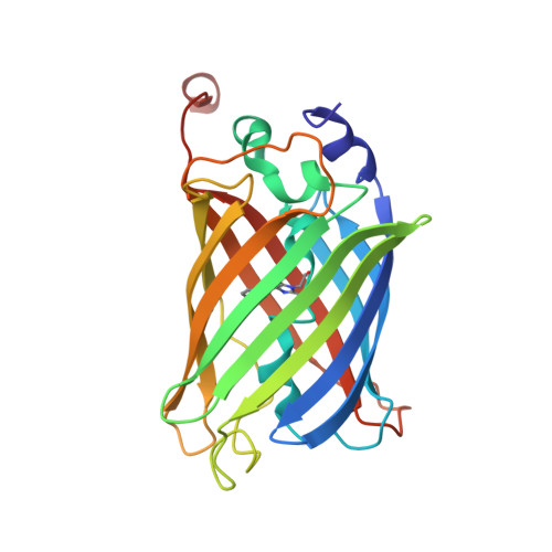
Structural basis for lipid and copper regulation of the ABC transporter MsbA.

Lyu, J.Liu, C.Zhang, T.Schrecke, S.Elam, N.P.Packianathan, C.Hochberg, G.K.A.Russell, D.Zhao, M.Laganowsky, A.

(2022) Nat Commun 13: 7291-7291

  • DOI: https://doi.org/10.1038/s41467-022-34905-2
  • Primary Citation Related Structures: 
    8DHY, 8DMM, 8DMO

  • PubMed Abstract: 

    A critical step in lipopolysaccharide (LPS) biogenesis involves flipping lipooligosaccharide, an LPS precursor, from the cytoplasmic to the periplasmic leaflet of the inner membrane, an operation carried out by the ATP-binding cassette transporter MsbA. Although LPS binding to the inner cavity of MsbA is well established, the selectivity of MsbA-lipid interactions at other site(s) remains poorly understood. Here we use native mass spectrometry (MS) to characterize MsbA-lipid interactions and guide structural studies. We show the transporter co-purifies with copper(II) and metal binding modulates protein-lipid interactions. A 2.15 Å resolution structure of an N-terminal region of MsbA in complex with copper(II) is presented, revealing a structure reminiscent of the GHK peptide, a high-affinity copper(II) chelator. Our results demonstrate conformation-dependent lipid binding affinities, particularly for the LPS-precursor, 3-deoxy-D-manno-oct-2-ulosonic acid (Kdo) 2 -lipid A (KDL). We report a 3.6 Å-resolution structure of MsbA trapped in an open, outward-facing conformation with adenosine 5'-diphosphate and vanadate, revealing a distinct KDL binding site, wherein the lipid forms extensive interactions with the transporter. Additional studies provide evidence that the exterior KDL binding site is conserved and a positive allosteric modulator of ATPase activity, serving as a feedforward activation mechanism to couple transporter activity with LPS biosynthesis.


  • Organizational Affiliation
    • Department of Chemistry, Texas A&M University, College Station, 77843, TX, USA.

Macromolecule Content 

  • Total Structure Weight: 27.24 kDa 
  • Atom Count: 1,941 
  • Modeled Residue Count: 237 
  • Deposited Residue Count: 239 
  • Unique protein chains: 1

Macromolecules

Find similar proteins by:|  3D Structure
Entity ID: 1
MoleculeChains  Sequence LengthOrganismDetailsImage
Fusion protein of MsbA N-terminal fragment and GFP,Green fluorescent protein239Escherichia coliAequorea victoria
This entity is chimeric
Mutation(s): 1 
Gene Names: msbAgfp
EC: 7.5.2.6
Membrane Entity: Yes 
UniProt
Find proteins for P60752 (Escherichia coli (strain K12))
Explore P60752 
Go to UniProtKB:  P60752
Entity Groups
Sequence Clusters30% Identity50% Identity70% Identity90% Identity95% Identity100% Identity
UniProt GroupP60752
Sequence Annotations
Expand
Reference Sequence

Small Molecules

Ligands 1 Unique
IDChains Name / Formula / InChI Key2D Diagram3D Interactions
CU
(Subject of Investigation/LOI)

Query on CU


Download:Ideal Coordinates CCD File
B [auth A]COPPER (II) ION
Cu
JPVYNHNXODAKFH-UHFFFAOYSA-N
Modified Residues  1 Unique
IDChains TypeFormula2D DiagramParent
CRO
Query on CRO
A
L-PEPTIDE LINKINGC15 H17 N3 O5THR, TYR, GLY

Experimental Data & Validation

Experimental Data

  • Method: X-RAY DIFFRACTION
  • Resolution: 2.15 Å
  • R-Value Free:  0.228 (Depositor), 0.232 (DCC) 
  • R-Value Work:  0.202 (Depositor), 0.209 (DCC) 
  • R-Value Observed: 0.203 (Depositor) 
Space Group: I 21 21 21
Unit Cell:
Length ( Å )Angle ( ˚ )
a = 70.61α = 90
b = 108.54β = 90
c = 140.4γ = 90
Software Package:
Software NamePurpose
PHENIXrefinement
XDSdata reduction
XSCALEdata scaling
Cootmodel building
PHENIXphasing

Structure Validation

View Full Validation Report


Entry History 

& Funding Information

Deposition Data


Funding OrganizationLocationGrant Number
National Institutes of Health/National Institute of General Medical Sciences (NIH/NIGMS)United StatesR01GM139876
National Institutes of Health/National Institute of General Medical Sciences (NIH/NIGMS)United StatesR01GM138863

Revision History  (Full details and data files)

  • Version 1.0: 2022-12-07
    Type: Initial release
  • Version 1.1: 2023-10-25
    Changes: Data collection, Refinement description
  • Version 1.2: 2023-11-15
    Changes: Data collection
  • Version 1.3: 2024-10-23
    Changes: Structure summary
  • Version 2.0: 2026-03-18
    Changes: Polymer sequence