8DAF

Human SF-1 LBD bound to synthetic agonist 6N-10CA and bacterial phospholipid


Experimental Data Snapshot

  • Method: X-RAY DIFFRACTION
  • Resolution: 2.59 Å
  • R-Value Free: 0.281 
  • R-Value Work: 0.223 
  • R-Value Observed: 0.226 

wwPDB Validation   3D Report Full Report


Ligand Structure Quality Assessment 


This is version 1.2 of the entry. See complete history


Literature

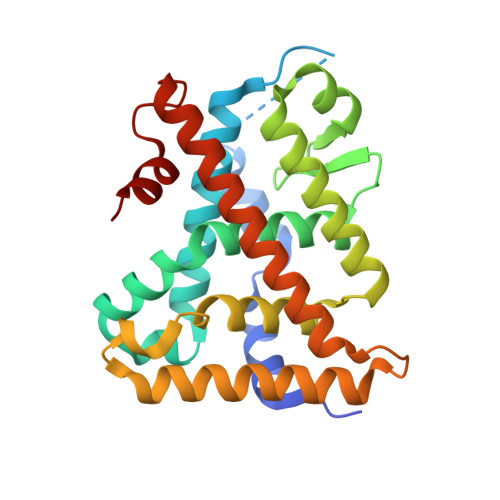
Comparison of activity, structure, and dynamics of SF-1 and LRH-1 complexed with small molecule modulators.

Cato, M.L.D'Agostino, E.H.Spurlin, R.M.Flynn, A.R.Cornelison, J.L.Johnson, A.M.Fujita, R.A.Abraham, S.M.Jui, N.T.Ortlund, E.A.

(2023) J Biol Chem 299: 104921-104921

  • DOI: https://doi.org/10.1016/j.jbc.2023.104921
  • Primary Citation of Related Structures:  
    8DAF

  • PubMed Abstract: 

    Steroidogenic factor-1 (SF-1) is a phospholipid-sensing nuclear receptor expressed in the adrenal glands, gonads, and hypothalamus which controls steroidogenesis and metabolism. There is significant therapeutic interest in SF-1 because of its oncogenic properties in adrenocortical cancer. Synthetic modulators are attractive for targeting SF-1 for clinical and laboratory purposes due to the poor pharmaceutical properties of its native phospholipid ligands. While small molecule agonists targeting SF-1 have been synthesized, no crystal structures have been reported of SF-1 in complexes with synthetic compounds. This has prevented the establishment of structure-activity relationships that would enable better characterization of ligand-mediated activation and improvement in current chemical scaffolds. Here, we compare the effects of small molecules in SF-1 and its close homolog, liver receptor homolog-1 (LRH-1), and identify several molecules that specifically activate LRH-1. We also report the first crystal structure of SF-1 in complex with a synthetic agonist that displays low nanomolar affinity and potency for SF-1. We use this structure to explore the mechanistic basis for small molecule agonism of SF-1, especially compared to LRH-1, and uncover unique signaling pathways that drive LRH-1 specificity. Molecular dynamics simulations reveal differences in protein dynamics at the pocket mouth as well as ligand-mediated allosteric communication from this region to the coactivator binding interface. Our studies, therefore, shed important insight into the allostery driving SF-1 activity and show potential for modulation of LRH-1 over SF-1.


  • Organizational Affiliation

    Department of Biochemistry, Emory University School of Medicine, Atlanta, Georgia, USA.


Macromolecules
Find similar proteins by:  (by identity cutoff)  |  3D Structure
Entity ID: 1
MoleculeChains Sequence LengthOrganismDetailsImage
Steroidogenic factor 1
A, B
247Homo sapiensMutation(s): 0 
Gene Names: NR5A1AD4BPFTZF1SF1
UniProt & NIH Common Fund Data Resources
Find proteins for Q13285 (Homo sapiens)
Explore Q13285 
Go to UniProtKB:  Q13285
PHAROS:  Q13285
GTEx:  ENSG00000136931 
Entity Groups  
Sequence Clusters30% Identity50% Identity70% Identity90% Identity95% Identity100% Identity
UniProt GroupQ13285
Sequence Annotations
Expand
  • Reference Sequence

Find similar proteins by:  Sequence   |   3D Structure  

Entity ID: 2
MoleculeChains Sequence LengthOrganismDetailsImage
Nuclear receptor coactivator 2
C, D
14Homo sapiensMutation(s): 0 
Gene Names: NCOA2BHLHE75SRC2TIF2
UniProt & NIH Common Fund Data Resources
Find proteins for Q15596 (Homo sapiens)
Explore Q15596 
Go to UniProtKB:  Q15596
PHAROS:  Q15596
GTEx:  ENSG00000140396 
Entity Groups  
Sequence Clusters30% Identity50% Identity70% Identity90% Identity95% Identity100% Identity
UniProt GroupQ15596
Sequence Annotations
Expand
  • Reference Sequence
Small Molecules
Ligands 2 Unique
IDChains Name / Formula / InChI Key2D Diagram3D Interactions
PEF
Query on PEF

Download Ideal Coordinates CCD File 
F [auth A],
H [auth B]
DI-PALMITOYL-3-SN-PHOSPHATIDYLETHANOLAMINE
C37 H74 N O8 P
SLKDGVPOSSLUAI-PGUFJCEWSA-N
IUW (Subject of Investigation/LOI)
Query on IUW

Download Ideal Coordinates CCD File 
E [auth A],
G [auth B]
10-[(3aR,6S,6aR)-3-phenyl-3a-(1-phenylethenyl)-6-(sulfamoylamino)-1,3a,4,5,6,6a-hexahydropentalen-2-yl]decanoic acid (non-preferred name)
C32 H42 N2 O4 S
SLIDCWKPFMEUQP-OLWNVYNHSA-N
Experimental Data & Validation

Experimental Data

  • Method: X-RAY DIFFRACTION
  • Resolution: 2.59 Å
  • R-Value Free: 0.281 
  • R-Value Work: 0.223 
  • R-Value Observed: 0.226 
  • Space Group: P 31 2 1
Unit Cell:
Length ( Å )Angle ( ˚ )
a = 73.428α = 90
b = 73.428β = 90
c = 194.184γ = 120
Software Package:
Software NamePurpose
PHENIXrefinement
HKL-2000data reduction
HKL-2000data scaling
PHASERphasing

Structure Validation

View Full Validation Report


Ligand Structure Quality Assessment 


Entry History & Funding Information

Deposition Data


Funding OrganizationLocationGrant Number
National Institutes of Health/National Institute of Diabetes and Digestive and Kidney Disease (NIH/NIDDK)United StatesF31DK122745
National Institutes of Health/National Institute of Diabetes and Digestive and Kidney Disease (NIH/NIDDK)United StatesR01DK115213
National Science Foundation (NSF, United States)United StatesDGE1444932

Revision History  (Full details and data files)

  • Version 1.0: 2023-06-28
    Type: Initial release
  • Version 1.1: 2023-08-09
    Changes: Database references
  • Version 1.2: 2023-10-25
    Changes: Data collection, Refinement description