8D3Z | pdb_00008d3z

Crystal structure of GalS1 in complex with Manganese from Populus trichocarpas


Experimental Data Snapshot

  • Method: X-RAY DIFFRACTION
  • Resolution: 2.56 Å
  • R-Value Free: 
    0.264 (Depositor), 0.264 (DCC) 
  • R-Value Work: 
    0.230 (Depositor), 0.230 (DCC) 
  • R-Value Observed: 
    0.231 (Depositor) 

Starting Model: experimental
View more details

wwPDB Validation 3D Report Full Report

Validation slider image for 8D3Z

Ligand Structure Quality Assessment 


This is version 1.3 of the entry. See complete history

Literature

Structural and biochemical insight into a modular beta-1,4-galactan synthase in plants.

Prabhakar, P.K.Pereira, J.H.Taujale, R.Shao, W.Bharadwaj, V.S.Chapla, D.Yang, J.Y.Bomble, Y.J.Moremen, K.W.Kannan, N.Hammel, M.Adams, P.D.Scheller, H.V.Urbanowicz, B.R.

(2023) Nat Plants 9: 486-500

  • DOI: https://doi.org/10.1038/s41477-023-01358-4
  • Primary Citation Related Structures: 
    8D3T, 8D3Z

  • PubMed Abstract: 

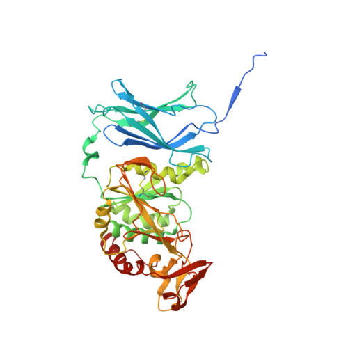
    Rhamnogalacturonan I (RGI) is a structurally complex pectic polysaccharide with a backbone of alternating rhamnose and galacturonic acid residues substituted with arabinan and galactan side chains. Galactan synthase 1 (GalS1) transfers galactose and arabinose to either extend or cap the β-1,4-galactan side chains of RGI, respectively. Here we report the structure of GalS1 from Populus trichocarpa, showing a modular protein consisting of an N-terminal domain that represents the founding member of a new family of carbohydrate-binding module, CBM95, and a C-terminal glycosyltransferase family 92 (GT92) catalytic domain that adopts a GT-A fold. GalS1 exists as a dimer in vitro, with stem domains interacting across the chains in a 'handshake' orientation that is essential for maintaining stability and activity. In addition to understanding the enzymatic mechanism of GalS1, we gained insight into the donor and acceptor substrate binding sites using deep evolutionary analysis, molecular simulations and biochemical studies. Combining all the results, a mechanism for GalS1 catalysis and a new model for pectic galactan side-chain addition are proposed.


  • Organizational Affiliation
    • Department of Biochemistry and Molecular Biology, University of Georgia, Athens, GA, USA.

Macromolecule Content 

  • Total Structure Weight: 102.89 kDa 
  • Atom Count: 6,928 
  • Modeled Residue Count: 798 
  • Deposited Residue Count: 848 
  • Unique protein chains: 1

Macromolecules

Find similar proteins by:|  3D Structure
Entity ID: 1
MoleculeChains  Sequence LengthOrganismDetailsImage
Galactan synthase
A, B
424Populus trichocarpaMutation(s): 0 
EC: 2.4.1
UniProt
Find proteins for A0A2K2AMS7 (Populus trichocarpa)
Explore A0A2K2AMS7 
Go to UniProtKB:  A0A2K2AMS7
Entity Groups
Sequence Clusters30% Identity50% Identity70% Identity90% Identity95% Identity100% Identity
UniProt GroupA0A2K2AMS7
Glycosylation
Glycosylation Sites: 4
Sequence Annotations
Expand
Reference Sequence

Oligosaccharides

Help  
Entity ID: 2
MoleculeChains Length2D Diagram GlycosylationD Interactions
2-acetamido-2-deoxy-beta-D-glucopyranose-(1-4)-2-acetamido-2-deoxy-beta-D-glucopyranose
C, E
2N-Glycosylation
Glycosylation Resources
GlyTouCan: G42666HT
GlyCosmos: G42666HT
GlyGen: G42666HT
Entity ID: 3
MoleculeChains Length2D Diagram GlycosylationD Interactions
alpha-D-mannopyranose-(1-2)-alpha-D-mannopyranose-(1-2)-alpha-D-mannopyranose-(1-3)-[alpha-D-mannopyranose-(1-6)]beta-D-mannopyranose-(1-4)-2-acetamido-2-deoxy-beta-D-glucopyranose-(1-4)-2-acetamido-2-deoxy-beta-D-glucopyranose
D, F
7N-Glycosylation
Glycosylation Resources
GlyTouCan: G46836GH
GlyCosmos: G46836GH
GlyGen: G46836GH

Small Molecules

Ligands 2 Unique
IDChains Name / Formula / InChI Key2D Diagram3D Interactions
NAG
(Subject of Investigation/LOI)

Query on NAG


Download:Ideal Coordinates CCD File
H [auth A]
I [auth A]
J [auth A]
K [auth A]
M [auth B]
H [auth A],
I [auth A],
J [auth A],
K [auth A],
M [auth B],
N [auth B],
O [auth B],
P [auth B]
2-acetamido-2-deoxy-beta-D-glucopyranose
C8 H15 N O6
OVRNDRQMDRJTHS-FMDGEEDCSA-N
MN

Query on MN


Download:Ideal Coordinates CCD File
G [auth A],
L [auth B]
MANGANESE (II) ION
Mn
WAEMQWOKJMHJLA-UHFFFAOYSA-N

Experimental Data & Validation

Experimental Data

  • Method: X-RAY DIFFRACTION
  • Resolution: 2.56 Å
  • R-Value Free:  0.264 (Depositor), 0.264 (DCC) 
  • R-Value Work:  0.230 (Depositor), 0.230 (DCC) 
  • R-Value Observed: 0.231 (Depositor) 
Space Group: P 31 2 1
Unit Cell:
Length ( Å )Angle ( ˚ )
a = 169.982α = 90
b = 169.982β = 90
c = 73.96γ = 120
Software Package:
Software NamePurpose
PHENIXrefinement
DIALSdata reduction
xia2data scaling
PHASERphasing

Structure Validation

View Full Validation Report


Ligand Structure Quality Assessment 


Entry History 

& Funding Information

Deposition Data


Funding OrganizationLocationGrant Number
Department of Energy (DOE, United States)United States--

Revision History  (Full details and data files)

  • Version 1.0: 2023-03-15
    Type: Initial release
  • Version 1.1: 2023-04-05
    Changes: Database references
  • Version 1.2: 2023-10-25
    Changes: Data collection, Refinement description
  • Version 1.3: 2024-10-30
    Changes: Structure summary