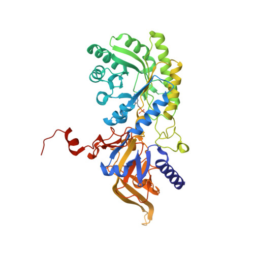
8D2Y

Y430F mutant of D-ornithine/D-lysine decarboxylase


Experimental Data Snapshot

  • Method: X-RAY DIFFRACTION
  • Resolution: 1.22 Å
  • R-Value Free: 0.152 
  • R-Value Work: 0.134 
  • R-Value Observed: 0.134 

wwPDB Validation   3D Report Full Report


This is version 1.2 of the entry. See complete history


Literature

The Y430F mutant of Salmonella d-ornithine/d-lysine decarboxylase has altered stereospecificity and a putrescine allosteric activation site.

Phillips, R.S.Nguyen Hoang, K.N.

(2022) Arch Biochem Biophys 731: 109429-109429

  • DOI: https://doi.org/10.1016/j.abb.2022.109429
  • Primary Citation of Related Structures:  
    8D2Y, 8D4I, 8D5D, 8D5R, 8D88

  • PubMed Abstract: 

    Tyrosine-430 of d-ornithine/d-lysine decarboxylase (DOKDC) is located in the active site, and was suggested to be responsible for the D-stereospecificity of the enzyme. We have prepared the Y430F mutant form of Salmonella enterica serovar typhimurium DOKDC and evaluated its catalytic activity with D- and l-lysine and ornithine. The kinetic results show that the Y430F mutant has measurable decarboxylase activity with both D- and l-lysine and ornithine, which wild type DOKDC does not. Spectroscopic experiments show that these amino acids bind to form external aldimine complexes with the pyridoxal-5'-phosphate with λ max  = 425 nm. In addition, we have obtained crystal structures of Y430F DOKDC bound to HEPES, putrescine, d-ornithine, d-lysine, and d-arginine. The d-amino acids bind in the crystals to form equilibrium mixtures of gem-diamine and external aldimine complexes. Furthermore, the crystal structures reveal an unexpected allosteric product activator site for putrescine located on the 2-fold axis between the two active sites. Putrescine binds by donating hydrogen bonds from the ammonium groups to Asp-361 and Gln-358 in the specificity helix of both chains. Addition of 0.1-1 mM putrescine eliminates the lag in steady state kinetics and abolishes the sigmoid kinetics. The catalytic loop was modeled with AlphaFold2, and the model shows that Glu-181 can form additional hydrogen bonds with the bound putrescine, likely stabilizing the catalytic closed conformation.


  • Organizational Affiliation

    Department of Chemistry, University of Georgia, Athens, GA, 30602, USA; Department of Biochemistry and Molecular Biology, University of Georgia, Athens, GA, 30602, USA. Electronic address: plp@uga.edu.


Macromolecules
Find similar proteins by:  (by identity cutoff)  |  3D Structure
Entity ID: 1
MoleculeChains Sequence LengthOrganismDetailsImage
D-ornithine/D-lysine decarboxylase477Salmonella enterica subsp. enterica serovar TyphimuriumMutation(s): 1 
Gene Names: dokDSTM2360
EC: 4.1.1.116
UniProt
Find proteins for Q8ZNC4 (Salmonella typhimurium (strain LT2 / SGSC1412 / ATCC 700720))
Explore Q8ZNC4 
Go to UniProtKB:  Q8ZNC4
Entity Groups  
Sequence Clusters30% Identity50% Identity70% Identity90% Identity95% Identity100% Identity
UniProt GroupQ8ZNC4
Sequence Annotations
Expand
  • Reference Sequence
Small Molecules
Ligands 7 Unique
IDChains Name / Formula / InChI Key2D Diagram3D Interactions
EPE
Query on EPE

Download Ideal Coordinates CCD File 
E [auth A]4-(2-HYDROXYETHYL)-1-PIPERAZINE ETHANESULFONIC ACID
C8 H18 N2 O4 S
JKMHFZQWWAIEOD-UHFFFAOYSA-N
GOL
Query on GOL

Download Ideal Coordinates CCD File 
J [auth A],
K [auth A]
GLYCEROL
C3 H8 O3
PEDCQBHIVMGVHV-UHFFFAOYSA-N
DMS
Query on DMS

Download Ideal Coordinates CCD File 
B [auth A],
D [auth A],
G [auth A]
DIMETHYL SULFOXIDE
C2 H6 O S
IAZDPXIOMUYVGZ-UHFFFAOYSA-N
EDO
Query on EDO

Download Ideal Coordinates CCD File 
C [auth A],
I [auth A]
1,2-ETHANEDIOL
C2 H6 O2
LYCAIKOWRPUZTN-UHFFFAOYSA-N
ACT
Query on ACT

Download Ideal Coordinates CCD File 
F [auth A],
H [auth A]
ACETATE ION
C2 H3 O2
QTBSBXVTEAMEQO-UHFFFAOYSA-M
K
Query on K

Download Ideal Coordinates CCD File 
M [auth A]POTASSIUM ION
K
NPYPAHLBTDXSSS-UHFFFAOYSA-N
CL
Query on CL

Download Ideal Coordinates CCD File 
L [auth A]CHLORIDE ION
Cl
VEXZGXHMUGYJMC-UHFFFAOYSA-M
Modified Residues  1 Unique
IDChains TypeFormula2D DiagramParent
LLP
Query on LLP
A
L-PEPTIDE LINKINGC14 H22 N3 O7 PLYS
Experimental Data & Validation

Experimental Data

  • Method: X-RAY DIFFRACTION
  • Resolution: 1.22 Å
  • R-Value Free: 0.152 
  • R-Value Work: 0.134 
  • R-Value Observed: 0.134 
  • Space Group: C 1 2 1
Unit Cell:
Length ( Å )Angle ( ˚ )
a = 139.74α = 90
b = 50.74β = 120.8
c = 73.32γ = 90
Software Package:
Software NamePurpose
PHENIXrefinement
autoPROCdata reduction
Aimlessdata scaling
PHASERphasing

Structure Validation

View Full Validation Report


Entry History & Funding Information

Deposition Data


Funding OrganizationLocationGrant Number
National Institutes of Health/National Institute of General Medical Sciences (NIH/NIGMS)United StatesR01 GM137008

Revision History  (Full details and data files)

  • Version 1.0: 2022-11-16
    Type: Initial release
  • Version 1.1: 2023-10-18
    Changes: Data collection, Refinement description
  • Version 1.2: 2023-11-15
    Changes: Data collection