8CWI | pdb_00008cwi

Fab arm of antibody 10G4 bound to CoV-2 receptor binding domain (RBD)


Experimental Data Snapshot

  • Method: X-RAY DIFFRACTION
  • Resolution: 1.87 Å
  • R-Value Free: 
    0.205 (Depositor), 0.206 (DCC) 
  • R-Value Work: 
    0.170 (Depositor), 0.172 (DCC) 
  • R-Value Observed: 
    0.172 (Depositor) 

Starting Model: experimental
View more details

wwPDB Validation 3D Report Full Report

Validation slider image for 8CWI

This is version 1.3 of the entry. See complete history

Literature

Affinity maturation endows potent activity onto class 6 SARS-CoV-2 broadly neutralizing antibodies.

Mazigi, O.Langley, D.B.Henry, J.Y.Burnett, D.L.Sobti, M.Walker, G.J.Rouet, R.Balachandran, H.Lenthall, H.Jackson, J.Ubiparipovic, S.Schofield, P.Brown, S.H.J.Schulz, S.R.Hoffmann, M.Pohlmann, S.Post, J.Martinello, M.Ahlenstiel, G.Kelleher, A.Rawlinson, W.D.Turville, S.G.Bull, R.A.Stewart, A.G.Jack, H.M.Goodnow, C.C.Christ, D.

(2025) Proc Natl Acad Sci U S A 122: e2417544121-e2417544121

  • DOI: https://doi.org/10.1073/pnas.2417544121
  • Primary Citation Related Structures: 
    8CWI, 8CWJ, 8CWK

  • PubMed Abstract: 

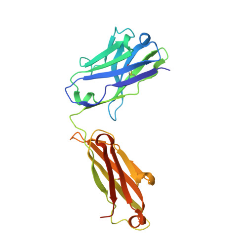
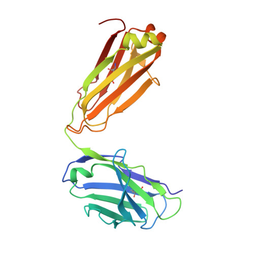
    The emergence of SARS-CoV-2 variants of concern (VOCs) has greatly diminished the neutralizing activity of previously FDA-approved monoclonal antibodies (mAbs), including that of antibody cocktails and of first-generation broadly neutralizing antibodies such as S309 (Sotrovimab). In contrast, antibodies targeting cryptic conformational epitopes of the receptor binding domain (RBD) have demonstrated broad activity against emerging variants, but exert only moderate neutralizing activity, which has so far hindered clinical development. Here, we utilize in vitro display technology to identify and affinity-mature antibodies targeting the cryptic class 6 epitope, accessible only in the "up" conformation of the SARS-CoV-2 spike trimer. Increasing antibody affinity into the low picomolar range endowed potent neutralization of VOCs and protection of hACE2 mice from viral challenge. Cryoelectron microscopy and crystal structures of two affinity-matured antibodies (4C12-B12 and 4G1-C2) in complex with RBD highlighted binding modes and epitopes distal from mutational hotspots commonly overserved in VOCs, providing direct structural insights into the observed mutational resistance. Moreover, we further demonstrate that antibodies targeting the class 6 epitope, rather than being an artifact of in vitro selection, are common in the IgG1 + memory B cell repertoire of convalescent patients and can be induced in human antibody V-gene transgenic mice through immunization. Our results highlight the importance of very high (picomolar) affinity in the development of neutralizing antibodies and vaccines and suggest an affinity threshold in the provision of broad and long-lasting immunity against SARS-CoV-2.


  • Organizational Affiliation
    • Garvan Institute of Medical Research, Sydney, NSW 2010, Australia.

Macromolecule Content 

  • Total Structure Weight: 72.82 kDa 
  • Atom Count: 5,666 
  • Modeled Residue Count: 627 
  • Deposited Residue Count: 647 
  • Unique protein chains: 3

Macromolecules

Find similar proteins by:|  3D Structure
Entity ID: 1
MoleculeChains  Sequence LengthOrganismDetailsImage
Spike protein S1204Severe acute respiratory syndrome coronavirus 2Mutation(s): 0 
Gene Names: S2
UniProt
Find proteins for P0DTC2 (Severe acute respiratory syndrome coronavirus 2)
Explore P0DTC2 
Go to UniProtKB:  P0DTC2
Entity Groups
Sequence Clusters30% Identity50% Identity70% Identity90% Identity95% Identity100% Identity
UniProt GroupP0DTC2
Glycosylation
Glycosylation Sites: 1Go to GlyGen: P0DTC2-1
Sequence Annotations
Expand
Reference Sequence
Find similar proteins by:|  3D Structure
Entity ID: 2
MoleculeChains  Sequence LengthOrganismDetailsImage
Heavy chain of Fab arm of antibody 10G4B [auth H]228Homo sapiensMutation(s): 0 
Find similar proteins by:|  3D Structure
Entity ID: 3
MoleculeChains  Sequence LengthOrganismDetailsImage
Light chain of Fab arm of antibody 10G4C [auth L]215Homo sapiensMutation(s): 0 

Oligosaccharides

Help  
Entity ID: 4
MoleculeChains Length2D Diagram GlycosylationD Interactions
alpha-D-mannopyranose-(1-3)-[alpha-D-mannopyranose-(1-6)]beta-D-mannopyranose-(1-4)-2-acetamido-2-deoxy-beta-D-glucopyranose-(1-4)-[alpha-L-fucopyranose-(1-6)]2-acetamido-2-deoxy-beta-D-glucopyranoseD [auth B]6N-Glycosylation
Glycosylation Resources
GlyTouCan: G82348BZ
GlyCosmos: G82348BZ
GlyGen: G82348BZ
Entity ID: 5
MoleculeChains Length2D Diagram GlycosylationD Interactions
2-acetamido-2-deoxy-beta-D-glucopyranose-(1-4)-[alpha-L-fucopyranose-(1-6)]2-acetamido-2-deoxy-beta-D-glucopyranoseE [auth C]3N-Glycosylation
Glycosylation Resources
GlyTouCan: G21290RB
GlyCosmos: G21290RB
GlyGen: G21290RB

Experimental Data & Validation

Experimental Data

  • Method: X-RAY DIFFRACTION
  • Resolution: 1.87 Å
  • R-Value Free:  0.205 (Depositor), 0.206 (DCC) 
  • R-Value Work:  0.170 (Depositor), 0.172 (DCC) 
  • R-Value Observed: 0.172 (Depositor) 
Space Group: P 1 21 1
Unit Cell:
Length ( Å )Angle ( ˚ )
a = 53.691α = 90
b = 88.639β = 95.03
c = 81.398γ = 90
Software Package:
Software NamePurpose
PHENIXrefinement
XDSdata reduction
Aimlessdata scaling
PHASERphasing
PDB_EXTRACTdata extraction

Structure Validation

View Full Validation Report


Entry History 

& Funding Information

Deposition Data


Funding OrganizationLocationGrant Number
National Health and Medical Research Council (NHMRC, Australia)Australia--

Revision History  (Full details and data files)

  • Version 1.0: 2023-05-24
    Type: Initial release
  • Version 1.1: 2024-04-03
    Changes: Data collection, Refinement description
  • Version 1.2: 2024-11-06
    Changes: Structure summary
  • Version 1.3: 2025-01-15
    Changes: Database references