8CU7

Crystal structure of A2AAR-StaR2-bRIL in complex with a novel A2a antagonist, LJ-4517


Experimental Data Snapshot

  • Method: X-RAY DIFFRACTION
  • Resolution: 2.05 Å
  • R-Value Free: 0.211 
  • R-Value Work: 0.177 
  • R-Value Observed: 0.179 

wwPDB Validation   3D Report Full Report


Ligand Structure Quality Assessment 


This is version 1.2 of the entry. See complete history


Literature

GPCR Agonist-to-Antagonist Conversion: Enabling the Design of Nucleoside Functional Switches for the A 2A Adenosine Receptor.

Shiriaeva, A.Park, D.Kim, G.Lee, Y.Hou, X.Jarhad, D.B.Kim, G.Yu, J.Hyun, Y.E.Kim, W.Gao, Z.G.Jacobson, K.A.Han, G.W.Stevens, R.C.Jeong, L.S.Choi, S.Cherezov, V.

(2022) J Med Chem 65: 11648-11657

  • DOI: https://doi.org/10.1021/acs.jmedchem.2c00462
  • Primary Citation of Related Structures:  
    8CU6, 8CU7

  • PubMed Abstract: 

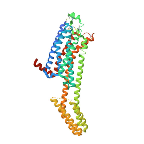
    Modulators of the G protein-coupled A 2A adenosine receptor (A 2A AR) have been considered promising agents to treat Parkinson's disease, inflammation, cancer, and central nervous system disorders. Herein, we demonstrate that a thiophene modification at the C8 position in the common adenine scaffold converted an A 2A AR agonist into an antagonist. We synthesized and characterized a novel A 2A AR antagonist, 2 (LJ-4517), with K i = 18.3 nM. X-ray crystallographic structures of 2 in complex with two thermostabilized A 2A AR constructs were solved at 2.05 and 2.80 Å resolutions. In contrast to A 2A AR agonists, which simultaneously interact with both Ser277 7.42 and His278 7.43 , 2 only transiently contacts His278 7.43 , which can be direct or water-mediated. The n -hexynyl group of 2 extends into an A 2A AR exosite. Structural analysis revealed that the introduced thiophene modification restricted receptor conformational rearrangements required for subsequent activation. This approach can expand the repertoire of adenosine receptor antagonists that can be designed based on available agonist scaffolds.


  • Organizational Affiliation

    Department of Chemistry, University of Southern California, Los Angeles, California 90089, United States.


Macromolecules
Find similar proteins by:  (by identity cutoff)  |  3D Structure
Entity ID: 1
MoleculeChains Sequence LengthOrganismDetailsImage
Adenosine receptor A2a,Soluble cytochrome b562447Homo sapiensEscherichia coliMutation(s): 12 
Gene Names: ADORA2AADORA2cybC
Membrane Entity: Yes 
UniProt & NIH Common Fund Data Resources
Find proteins for P0ABE7 (Escherichia coli)
Explore P0ABE7 
Go to UniProtKB:  P0ABE7
Find proteins for P29274 (Homo sapiens)
Explore P29274 
Go to UniProtKB:  P29274
PHAROS:  P29274
GTEx:  ENSG00000128271 
Entity Groups  
Sequence Clusters30% Identity50% Identity70% Identity90% Identity95% Identity100% Identity
UniProt GroupsP0ABE7P29274
Sequence Annotations
Expand
  • Reference Sequence
Small Molecules
Ligands 7 Unique
IDChains Name / Formula / InChI Key2D Diagram3D Interactions
LJX (Subject of Investigation/LOI)
Query on LJX

Download Ideal Coordinates CCD File 
C [auth A](2R,3R,4R)-2-[(8P)-6-amino-2-(hex-1-yn-1-yl)-8-(thiophen-2-yl)-9H-purin-9-yl]oxolane-3,4-diol
C19 H21 N5 O3 S
MUHXGASCCALSAJ-PRIMNGNOSA-N
CLR
Query on CLR

Download Ideal Coordinates CCD File 
J [auth A],
K [auth A],
L [auth A]
CHOLESTEROL
C27 H46 O
HVYWMOMLDIMFJA-DPAQBDIFSA-N
OLC
Query on OLC

Download Ideal Coordinates CCD File 
E [auth A]
G [auth A]
MA [auth A]
NA [auth A]
OA [auth A]
E [auth A],
G [auth A],
MA [auth A],
NA [auth A],
OA [auth A],
W [auth A],
X [auth A]
(2R)-2,3-dihydroxypropyl (9Z)-octadec-9-enoate
C21 H40 O4
RZRNAYUHWVFMIP-GDCKJWNLSA-N
OLA
Query on OLA

Download Ideal Coordinates CCD File 
AA [auth A]
BA [auth A]
CA [auth A]
DA [auth A]
EA [auth A]
AA [auth A],
BA [auth A],
CA [auth A],
DA [auth A],
EA [auth A],
F [auth A],
FA [auth A],
GA [auth A],
H [auth A],
HA [auth A],
I [auth A],
M [auth A],
N [auth A],
O [auth A],
P [auth A],
Q [auth A],
R [auth A],
S [auth A],
T [auth A],
U [auth A],
V [auth A],
Y [auth A],
Z [auth A]
OLEIC ACID
C18 H34 O2
ZQPPMHVWECSIRJ-KTKRTIGZSA-N
PEG
Query on PEG

Download Ideal Coordinates CCD File 
B [auth A],
JA [auth A],
KA [auth A],
LA [auth A]
DI(HYDROXYETHYL)ETHER
C4 H10 O3
MTHSVFCYNBDYFN-UHFFFAOYSA-N
ETF
Query on ETF

Download Ideal Coordinates CCD File 
IA [auth A]TRIFLUOROETHANOL
C2 H3 F3 O
RHQDFWAXVIIEBN-UHFFFAOYSA-N
NA
Query on NA

Download Ideal Coordinates CCD File 
D [auth A]SODIUM ION
Na
FKNQFGJONOIPTF-UHFFFAOYSA-N
Experimental Data & Validation

Experimental Data

  • Method: X-RAY DIFFRACTION
  • Resolution: 2.05 Å
  • R-Value Free: 0.211 
  • R-Value Work: 0.177 
  • R-Value Observed: 0.179 
  • Space Group: C 2 2 21
Unit Cell:
Length ( Å )Angle ( ˚ )
a = 39.456α = 90
b = 179.58β = 90
c = 140.873γ = 90
Software Package:
Software NamePurpose
PHENIXrefinement
PDB_EXTRACTdata extraction
HKL-2000data reduction
HKL-2000data scaling
PHASERphasing

Structure Validation

View Full Validation Report


Ligand Structure Quality Assessment 


Entry History & Funding Information

Deposition Data


Funding OrganizationLocationGrant Number
National Institutes of Health/National Institute of Diabetes and Digestive and Kidney Disease (NIH/NIDDK)United StatesDK31117

Revision History  (Full details and data files)

  • Version 1.0: 2022-08-31
    Type: Initial release
  • Version 1.1: 2022-09-21
    Changes: Database references
  • Version 1.2: 2023-10-18
    Changes: Data collection, Refinement description