8CU0

12-mer DNA structure of ExBIM bound to RNaseH -modified DDD


Experimental Data Snapshot

  • Method: X-RAY DIFFRACTION
  • Resolution: 1.74 Å
  • R-Value Free: 0.246 
  • R-Value Work: 0.202 
  • R-Value Observed: 0.204 

wwPDB Validation   3D Report Full Report


Ligand Structure Quality Assessment 


This is version 1.2 of the entry. See complete history


Literature

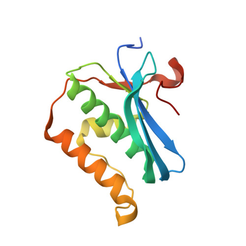
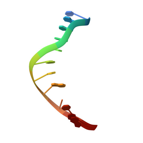
Conformation and Pairing Properties of an O 6 -Methyl-2'-deoxyguanosine-Directed Benzimidazole Nucleoside Analog in Duplex DNA.

Kellum Jr., A.H.Pallan, P.S.Nilforoushan, A.Sturla, S.J.Stone, M.P.Egli, M.

(2022) Chem Res Toxicol 35: 1903-1913

  • DOI: https://doi.org/10.1021/acs.chemrestox.2c00165
  • Primary Citation of Related Structures:  
    8CTY, 8CTZ, 8CU0

  • PubMed Abstract: 

    O 6 -Methyl-2'-deoxyguanosine ( O 6 -MeG) is one of the most common DNA lesions and arises as a consequence of both xenobiotic carcinogens and endogenous methylation by S -adenosylmethionine. O 6 -MeG frequently causes G-to-A mutations during DNA replication due to the misincorporation of dTTP and continued DNA synthesis. Efforts to detect DNA adducts such as O 6 -MeG, and to understand their impacts on DNA structure and function, have motivated the creation of nucleoside analogs with altered base moieties to afford a more favorable interaction with the adduct as compared to the unmodified nucleotide. Such analogs directed at O 6 -MeG include benzimidazolinone and benzimidazole nucleotides, as well as their extended π surface analogs naphthimidazolinone and napthimidazole derivatives. These analogs form a more stable pair with O 6 -MeG than with G, most likely due to a combination of H-bonding and stacking. While extending the π surface of the analogs enhances their performance as adduct-directed probes, the precise origins of the increased affinity between the synthetic analogs and O 6 -MeG remain unclear. To better understand relevant conformational and pairing properties, we used X-ray crystallography and analyzed the structures of the DNA duplexes with naphthimidazolinone inserted opposite G or O 6 -MeG. The structures reveal a complex interaction of the analog found either in an anti orientation and stacked inside the duplex, either above or below G or O 6 -MeG, or in a syn orientation and paired opposite G with formation of a single H-bond. The experimental structural data are consistent with the stabilizing effect of the synthetic analog observed in UV melting experiments and calculations and moreover reveal that the origin of these observations appears to be superior stacking between O 6 -MeG and the extended π system of the synthetic probe.


  • Organizational Affiliation

    Department of Chemistry, Vanderbilt University, College of Arts and Science, Nashville, Tennessee 37235, United States.


Macromolecules

Find similar proteins by:  (by identity cutoff)  |  3D Structure
Entity ID: 1
MoleculeChains Sequence LengthOrganismDetailsImage
Ribonuclease H
A, B, C
142Halalkalibacterium haloduransMutation(s): 1 
Gene Names: rnhA
EC: 3.1.26.4
UniProt
Find proteins for Q9KEI9 (Halalkalibacterium halodurans (strain ATCC BAA-125 / DSM 18197 / FERM 7344 / JCM 9153 / C-125))
Explore Q9KEI9 
Go to UniProtKB:  Q9KEI9
Entity Groups  
Sequence Clusters30% Identity50% Identity70% Identity90% Identity95% Identity100% Identity
UniProt GroupQ9KEI9
Sequence Annotations
Expand
  • Reference Sequence

Find similar nucleic acids by:  Sequence   |   3D Structure  

Entity ID: 2
MoleculeChains LengthOrganismImage
DNA (5'-D(*CP*GP*GP*GP*CP*AP*TP*GP*(OWR)P*CP*CP*G)-3')
D, E
12synthetic construct
Sequence Annotations
Expand
  • Reference Sequence
Small Molecules
Ligands 3 Unique
IDChains Name / Formula / InChI Key2D Diagram3D Interactions
GOL (Subject of Investigation/LOI)
Query on GOL

Download Ideal Coordinates CCD File 
G [auth A],
L [auth B],
P [auth C]
GLYCEROL
C3 H8 O3
PEDCQBHIVMGVHV-UHFFFAOYSA-N
EDO (Subject of Investigation/LOI)
Query on EDO

Download Ideal Coordinates CCD File 
F [auth A],
J [auth B],
K [auth B],
N [auth C],
O [auth C]
1,2-ETHANEDIOL
C2 H6 O2
LYCAIKOWRPUZTN-UHFFFAOYSA-N
NA (Subject of Investigation/LOI)
Query on NA

Download Ideal Coordinates CCD File 
H [auth A],
I [auth A],
M [auth B],
Q [auth C]
SODIUM ION
Na
FKNQFGJONOIPTF-UHFFFAOYSA-N
Experimental Data & Validation

Experimental Data

  • Method: X-RAY DIFFRACTION
  • Resolution: 1.74 Å
  • R-Value Free: 0.246 
  • R-Value Work: 0.202 
  • R-Value Observed: 0.204 
  • Space Group: P 1 21 1
Unit Cell:
Length ( Å )Angle ( ˚ )
a = 36.894α = 90
b = 89.403β = 101.42
c = 73.89γ = 90
Software Package:
Software NamePurpose
PHENIXrefinement
HKL-2000data reduction
HKL-2000data scaling
MOLREPphasing

Structure Validation

View Full Validation Report


Ligand Structure Quality Assessment 


Entry History & Funding Information

Deposition Data


Funding OrganizationLocationGrant Number
National Institutes of Health/National Cancer Institute (NIH/NCI)United StatesCA160032

Revision History  (Full details and data files)

  • Version 1.0: 2022-08-31
    Type: Initial release
  • Version 1.1: 2022-11-02
    Changes: Database references
  • Version 1.2: 2023-10-18
    Changes: Data collection, Refinement description