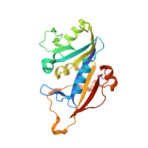
8CRH

Crystal structure of Candida auris dihydrofolate reductase complexed with NADPH and cycloguanil


Experimental Data Snapshot

  • Method: X-RAY DIFFRACTION
  • Resolution: 1.30 Å
  • R-Value Free: 0.183 
  • R-Value Work: 0.173 
  • R-Value Observed: 0.174 

wwPDB Validation   3D Report Full Report


Ligand Structure Quality Assessment 


This is version 1.1 of the entry. See complete history


Literature

Crystal structure of dihydrofolate reductase from the emerging pathogenic fungus Candida auris.

Kirkman, T.Sketcher, A.de Morais Barroso, V.Ishida, K.Tosin, M.Dias, M.V.B.

(2023) Acta Crystallogr D Struct Biol 79: 735-745

  • DOI: https://doi.org/10.1107/S2059798323004709
  • Primary Citation of Related Structures:  
    7ZZX, 8CRH

  • PubMed Abstract: 

    Candida auris has emerged as a global health problem with a dramatic spread by nosocomial transmission and a high mortality rate. Antifungal therapy for C. auris infections is currently limited due to widespread resistance to fluconazole and amphotericin B and increasing resistance to the front-line drug echinocandin. Therefore, new treatments are urgently required to combat this pathogen. Dihydrofolate reductase (DHFR) has been validated as a potential drug target for Candida species, although no structure of the C. auris enzyme (CauDHFR) has been reported. Here, crystal structures of CauDHFR are reported as an apoenzyme, as a holoenzyme and in two ternary complexes with pyrimethamine and cycloguanil, which are common antifolates, at near-atomic resolution. Preliminary biochemical and biophysical assays and antifungal susceptibility testing with a variety of classical antifolates were also performed, highlighting the enzyme-inhibition rates and the inhibition of yeast growth. These structural and functional data might provide the basis for a novel drug-discovery campaign against this global threat.


  • Organizational Affiliation

    Department of Chemistry, University of Warwick, Coventry CV4 7AL, United Kingdom.


Macromolecules
Find similar proteins by:  (by identity cutoff)  |  3D Structure
Entity ID: 1
MoleculeChains Sequence LengthOrganismDetailsImage
Dihydrofolate reductaseA [auth B],
B [auth A]
208[Candida] aurisMutation(s): 0 
Gene Names: QG37_02791
UniProt
Find proteins for A0A0L0P1H8 (Candida auris)
Explore A0A0L0P1H8 
Go to UniProtKB:  A0A0L0P1H8
Entity Groups  
Sequence Clusters30% Identity50% Identity70% Identity90% Identity95% Identity100% Identity
UniProt GroupA0A0L0P1H8
Sequence Annotations
Expand
  • Reference Sequence
Small Molecules
Ligands 3 Unique
IDChains Name / Formula / InChI Key2D Diagram3D Interactions
NDP (Subject of Investigation/LOI)
Query on NDP

Download Ideal Coordinates CCD File 
D [auth B],
H [auth A]
NADPH DIHYDRO-NICOTINAMIDE-ADENINE-DINUCLEOTIDE PHOSPHATE
C21 H30 N7 O17 P3
ACFIXJIJDZMPPO-NNYOXOHSSA-N
VIL (Subject of Investigation/LOI)
Query on VIL

Download Ideal Coordinates CCD File 
C [auth B],
E [auth B],
G [auth A],
I [auth A]
5-(4-chlorophenyl)-6,6-dimethyl-1,4-dihydro-1,3,5-triazine-2,4-diamine
C11 H16 Cl N5
NDMGACCSLHFEAQ-SNVBAGLBSA-N
SO4
Query on SO4

Download Ideal Coordinates CCD File 
F [auth B],
J [auth A],
K [auth A],
L [auth A],
M [auth A]
SULFATE ION
O4 S
QAOWNCQODCNURD-UHFFFAOYSA-L
Experimental Data & Validation

Experimental Data

  • Method: X-RAY DIFFRACTION
  • Resolution: 1.30 Å
  • R-Value Free: 0.183 
  • R-Value Work: 0.173 
  • R-Value Observed: 0.174 
  • Space Group: P 21 21 21
Unit Cell:
Length ( Å )Angle ( ˚ )
a = 43.776α = 90
b = 63.624β = 90
c = 167.583γ = 90
Software Package:
Software NamePurpose
Aimlessdata scaling
PHENIXrefinement
XDSdata reduction
PHASERphasing

Structure Validation

View Full Validation Report


Ligand Structure Quality Assessment 


Entry History & Funding Information

Deposition Data


Funding OrganizationLocationGrant Number
UK Research and Innovation (UKRI)United Kingdom--

Revision History  (Full details and data files)

  • Version 1.0: 2023-08-02
    Type: Initial release
  • Version 1.1: 2023-08-09
    Changes: Database references