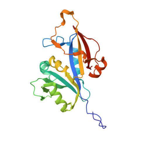
Crystal structure of dihydrofolate reductase from the emerging pathogenic fungus Candida auris.
Kirkman, T., Sketcher, A., de Morais Barroso, V., Ishida, K., Tosin, M., Dias, M.V.B.(2023) Acta Crystallogr D Struct Biol 79: 735-745
- PubMed: 37428844 Search on PubMedSearch on PubMed Central
- DOI: https://doi.org/10.1107/S2059798323004709
- Primary Citation Related Structures:
7ZZX, 8A0N, 8CRH - PubMed Abstract:
Candida auris has emerged as a global health problem with a dramatic spread by nosocomial transmission and a high mortality rate. Antifungal therapy for C. auris infections is currently limited due to widespread resistance to fluconazole and amphotericin B and increasing resistance to the front-line drug echinocandin. Therefore, new treatments are urgently required to combat this pathogen. Dihydrofolate reductase (DHFR) has been validated as a potential drug target for Candida species, although no structure of the C. auris enzyme (CauDHFR) has been reported. Here, crystal structures of CauDHFR are reported as an apoenzyme, as a holoenzyme and in two ternary complexes with pyrimethamine and cycloguanil, which are common antifolates, at near-atomic resolution. Preliminary biochemical and biophysical assays and antifungal susceptibility testing with a variety of classical antifolates were also performed, highlighting the enzyme-inhibition rates and the inhibition of yeast growth. These structural and functional data might provide the basis for a novel drug-discovery campaign against this global threat.
- Department of Chemistry, University of Warwick, Coventry CV4 7AL, United Kingdom.
Organizational Affiliation: