8CQY | pdb_00008cqy

Crystal structure of the PTPN3 PDZ domain bound to the PBM TACE C-terminal peptide


Experimental Data Snapshot

  • Method: X-RAY DIFFRACTION
  • Resolution: 1.70 Å
  • R-Value Free: 
    0.203 (Depositor), 0.195 (DCC) 
  • R-Value Work: 
    0.168 (Depositor), 0.155 (DCC) 
  • R-Value Observed: 
    0.169 (Depositor) 

Starting Model: experimental
View more details

wwPDB Validation 3D Report Full Report

Validation slider image for 8CQY

This is version 1.2 of the entry. See complete history

Literature

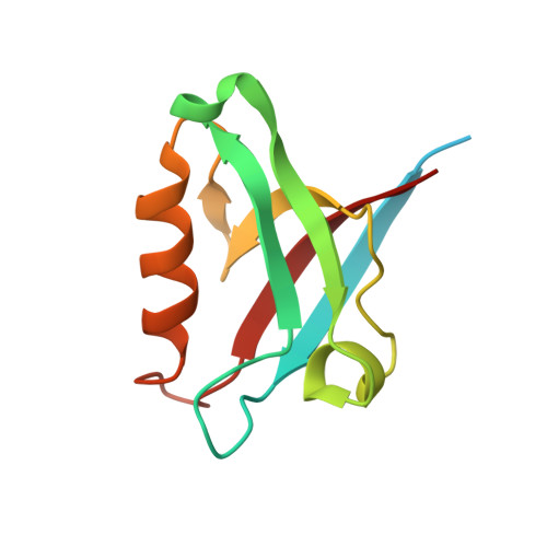
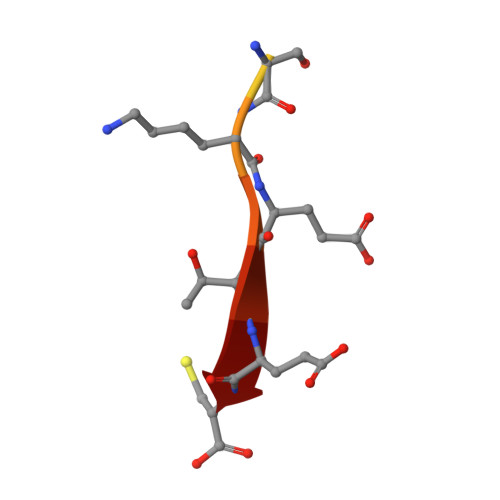
Interactions of the protein tyrosine phosphatase PTPN3 with viral and cellular partners through its PDZ domain: insights into structural determinants and phosphatase activity.

Genera, M.Colcombet-Cazenave, B.Croitoru, A.Raynal, B.Mechaly, A.Caillet, J.Haouz, A.Wolff, N.Caillet-Saguy, C.

(2023) Front Mol Biosci 10: 1192621-1192621

  • DOI: https://doi.org/10.3389/fmolb.2023.1192621
  • Primary Citation Related Structures: 
    8CQY, 8OEP

  • PubMed Abstract: 

    The human protein tyrosine phosphatase non-receptor type 3 (PTPN3) is a phosphatase containing a PDZ (PSD-95/Dlg/ZO-1) domain that has been found to play both tumor-suppressive and tumor-promoting roles in various cancers, despite limited knowledge of its cellular partners and signaling functions. Notably, the high-risk genital human papillomavirus (HPV) types 16 and 18 and the hepatitis B virus (HBV) target the PDZ domain of PTPN3 through PDZ-binding motifs (PBMs) in their E6 and HBc proteins respectively. This study focuses on the interactions between the PTPN3 PDZ domain (PTPN3-PDZ) and PBMs of viral and cellular protein partners. We solved the X-ray structures of complexes between PTPN3-PDZ and PBMs of E6 of HPV18 and the tumor necrosis factor-alpha converting enzyme (TACE). We provide new insights into key structural determinants of PBM recognition by PTPN3 by screening the selectivity of PTPN3-PDZ recognition of PBMs, and by comparing the PDZome binding profiles of PTPN3-recognized PBMs and the interactome of PTPN3-PDZ. The PDZ domain of PTPN3 was known to auto-inhibit the protein's phosphatase activity. We discovered that the linker connecting the PDZ and phosphatase domains is involved in this inhibition, and that the binding of PBMs does not impact this catalytic regulation. Overall, the study sheds light on the interactions and structural determinants of PTPN3 with its cellular and viral partners, as well as on the inhibitory role of its PDZ domain on its phosphatase activity.


  • Organizational Affiliation
    • Institut Pasteur, Université Paris Cité, Channel Receptors Unit, Paris, France.

Macromolecule Content 

  • Total Structure Weight: 14.22 kDa 
  • Atom Count: 808 
  • Modeled Residue Count: 99 
  • Deposited Residue Count: 126 
  • Unique protein chains: 2

Macromolecules

Find similar proteins by:|  3D Structure
Entity ID: 1
MoleculeChains  Sequence LengthOrganismDetailsImage
Tyrosine-protein phosphatase non-receptor type 3114Homo sapiensMutation(s): 0 
Gene Names: PTPN3PTPH1
EC: 3.1.3.48
UniProt & NIH Common Fund Data Resources
Find proteins for P26045 (Homo sapiens)
Explore P26045 
Go to UniProtKB:  P26045
PHAROS:  P26045
GTEx:  ENSG00000070159 
Entity Groups
Sequence Clusters30% Identity50% Identity70% Identity90% Identity95% Identity100% Identity
UniProt GroupP26045
Sequence Annotations
Expand
Reference Sequence
Find similar proteins by:  Sequence   |   3D Structure  
Entity ID: 2
MoleculeChains  Sequence LengthOrganismDetailsImage
Disintegrin and metalloproteinase domain-containing protein 1712Homo sapiensMutation(s): 0 
EC: 3.4.24.86
UniProt & NIH Common Fund Data Resources
Find proteins for P78536 (Homo sapiens)
Explore P78536 
Go to UniProtKB:  P78536
PHAROS:  P78536
GTEx:  ENSG00000151694 
Entity Groups
Sequence Clusters30% Identity50% Identity70% Identity90% Identity95% Identity100% Identity
UniProt GroupP78536
Sequence Annotations
Expand
Reference Sequence

Experimental Data & Validation

Experimental Data

  • Method: X-RAY DIFFRACTION
  • Resolution: 1.70 Å
  • R-Value Free:  0.203 (Depositor), 0.195 (DCC) 
  • R-Value Work:  0.168 (Depositor), 0.155 (DCC) 
  • R-Value Observed: 0.169 (Depositor) 
Space Group: P 32 2 1
Unit Cell:
Length ( Å )Angle ( ˚ )
a = 76.878α = 90
b = 76.878β = 90
c = 46.253γ = 120
Software Package:
Software NamePurpose
REFMACrefinement
XDSdata reduction
Aimlessdata scaling
PHASERphasing

Structure Validation

View Full Validation Report


Entry History 

& Funding Information

Deposition Data


Funding OrganizationLocationGrant Number
Pasteur InstituteFrance--
Centre National de la Recherche Scientifique (CNRS)France--

Revision History  (Full details and data files)

  • Version 1.0: 2023-05-10
    Type: Initial release
  • Version 1.1: 2023-05-31
    Changes: Database references
  • Version 1.2: 2024-06-19
    Changes: Data collection