8CL5 | pdb_00008cl5

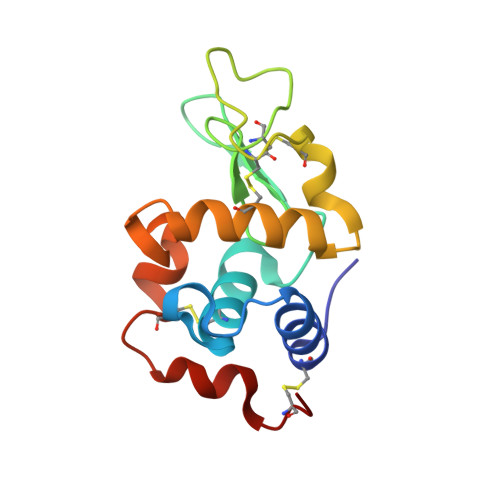
Lysozyme in matrix of lipidic cubic phase of monoolein (LCP)


Experimental Data Snapshot

  • Method: X-RAY DIFFRACTION
  • Resolution: 1.31 Å
  • R-Value Free: 
    0.148 (Depositor), 0.151 (DCC) 
  • R-Value Work: 
    0.125 (Depositor), 0.129 (DCC) 
  • R-Value Observed: 
    0.126 (Depositor) 

Starting Model: experimental
View more details

wwPDB Validation 3D Report Full Report

Validation slider image for 8CL5

This is version 1.1 of the entry. See complete history. 

Literature

A multi-reservoir extruder for time-resolved serial protein crystallography and compound screening at X-ray free-electron lasers.

Wranik, M., Kepa, M.W., Beale, E.V., James, D., Bertrand, Q., Weinert, T., Furrer, A., Glover, H., Gashi, D., Carrillo, M., Kondo, Y., Stipp, R.T., Khusainov, G., Nass, K., Ozerov, D., Cirelli, C., Johnson, P.J.M., Dworkowski, F., Beale, J.H., Stubbs, S., Zamofing, T., Schneider, M., Krauskopf, K., Gao, L., Thorn-Seshold, O., Bostedt, C., Bacellar, C., Steinmetz, M.O., Milne, C., Standfuss, J.

(2023) Nat Commun 14: 7956-7956

  • DOI: https://doi.org/10.1038/s41467-023-43523-5
  • Primary Citation Related Structures: 
    8CL5, 8CL6, 8CL7, 8CL8, 8CL9, 8CLB, 8CLC, 8CLD, 8CLE, 8CLF, 8CLG, 8CLH

  • PubMed Abstract: 

    Serial crystallography at X-ray free-electron lasers (XFELs) permits the determination of radiation-damage free static as well as time-resolved protein structures at room temperature. Efficient sample delivery is a key factor for such experiments. Here, we describe a multi-reservoir, high viscosity extruder as a step towards automation of sample delivery at XFELs. Compared to a standard single extruder, sample exchange time was halved and the workload of users was greatly reduced. In-built temperature control of samples facilitated optimal extrusion and supported sample stability. After commissioning the device with lysozyme crystals, we collected time-resolved data using crystals of a membrane-bound, light-driven sodium pump. Static data were also collected from the soluble protein tubulin that was soaked with a series of small molecule drugs. Using these data, we identify low occupancy (as little as 30%) ligands using a minimal amount of data from a serial crystallography experiment, a result that could be exploited for structure-based drug design.


  • Organizational Affiliation: 
    • Laboratory of Biomolecular Research, Division of Biology and Chemistry, Paul Scherrer Institut, Villigen-PSI, Villigen, 5232, Switzerland. maximilian.wranik@psi.ch.

Macromolecule Content 

  • Total Structure Weight: 14.75 kDa 
  • Atom Count: 1,329 
  • Modeled Residue Count: 129 
  • Deposited Residue Count: 129 
  • Unique protein chains: 1

Macromolecules

Find similar proteins by:|  3D Structure
Entity ID: 1
MoleculeChains  Sequence LengthOrganismDetailsImage
LYSOZYME, Chain A129Asplenium bulbiferum subsp. bulbiferumMutation(s): 0 
EC: 3.2.1.17
UniProt
Find proteins for P00698 (Gallus gallus)
Explore P00698 
Go to UniProtKB:  P00698
Entity Groups
Sequence Clusters30% Identity50% Identity70% Identity90% Identity95% Identity100% Identity
UniProt GroupP00698
Sequence Annotations
Expand
Reference Sequence

Small Molecules

Ligands 4 Unique
IDChains Name / Formula / InChI Key2D Diagram3D Interactions
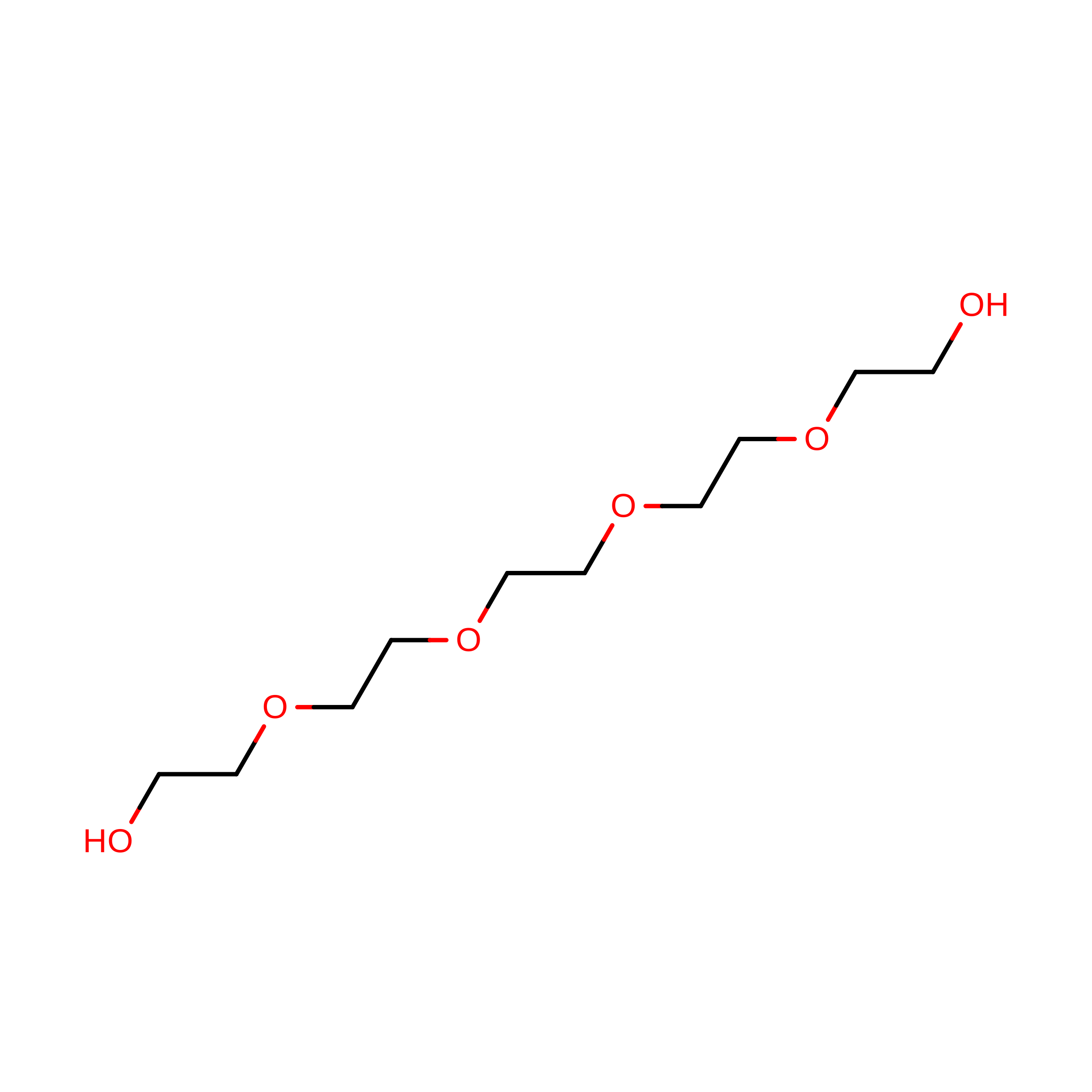
1PE

Query on 1PE


Download:Ideal Coordinates CCD File
G [auth A]PENTAETHYLENE GLYCOL
C10 H22 O6
JLFNLZLINWHATN-UHFFFAOYSA-N
EDO

Query on EDO


Download:Ideal Coordinates CCD File
B [auth A]1,2-ETHANEDIOL
C2 H6 O2
LYCAIKOWRPUZTN-UHFFFAOYSA-N
CL

Query on CL


Download:Ideal Coordinates CCD File
C [auth A],
D [auth A]
CHLORIDE ION
Cl
VEXZGXHMUGYJMC-UHFFFAOYSA-M
NA

Query on NA


Download:Ideal Coordinates CCD File
E [auth A],
F [auth A]
SODIUM ION
Na
FKNQFGJONOIPTF-UHFFFAOYSA-N

Experimental Data & Validation

Experimental Data

  • Method: X-RAY DIFFRACTION
  • Resolution: 1.31 Å
  • R-Value Free:  0.148 (Depositor), 0.151 (DCC) 
  • R-Value Work:  0.125 (Depositor), 0.129 (DCC) 
  • R-Value Observed: 0.126 (Depositor) 
Space Group: P 43 21 2
Unit Cell:
Length ( Å )Angle ( ˚ )
a = 77.92α = 90
b = 77.92β = 90
c = 37.64γ = 90
Software Package:
Software NamePurpose
PHENIXrefinement
CrystFELdata reduction
CrystFELdata scaling
PHASERphasing

Structure Validation

View Full Validation Report


Entry History 

& Funding Information

Deposition Data


Funding OrganizationLocationGrant Number
Swiss National Science FoundationSwitzerland31003A_179351
Swiss National Science FoundationSwitzerland310030_207462
Swiss National Science FoundationSwitzerland310030_192566

Revision History  (Full details and data files)

  • Version 1.0: 2023-12-13
    Type: Initial release
  • Version 1.1: 2024-11-20
    Changes: Structure summary