


Structural perspective on the design of selective DYRK1B inhibitors
Grygier, P., Pustelny, K., Dubin, G., Czarna, A.To be published.
Experimental Data Snapshot
Starting Model: experimental
View more details
Entity ID: 1 | |||||
|---|---|---|---|---|---|
| Molecule | Chains | Sequence Length | Organism | Details | Image |
| Dual specificity tyrosine-phosphorylation-regulated kinase 1A | 371 | Homo sapiens | Mutation(s): 0 Gene Names: DYRK1A, DYRK, MNB, MNBH EC: 2.7.11.23 (PDB Primary Data), 2.7.12.1 (PDB Primary Data) | 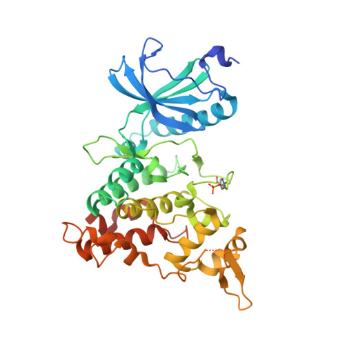 | |
UniProt & NIH Common Fund Data Resources | |||||
PHAROS: Q13627 GTEx: ENSG00000157540 | |||||
Entity Groups | |||||
| Sequence Clusters | 30% Identity50% Identity70% Identity90% Identity95% Identity100% Identity | ||||
| UniProt Group | Q13627 | ||||
Sequence AnnotationsExpand | |||||
Reference Sequence | |||||
| Ligands 3 Unique | |||||
|---|---|---|---|---|---|
| ID | Chains | Name / Formula / InChI Key | 2D Diagram | 3D Interactions | |
| QS0 (Subject of Investigation/LOI) Download:Ideal Coordinates CCD File | AA [auth C], E [auth A], JA [auth D], T [auth B] | N-[2-methoxy-4-(4-methylpiperazin-1-yl)phenyl]-4-(1-methylpyrrolo[2,3-c]pyridin-3-yl)pyrimidin-2-amine C24 H27 N7 O ZYVXTMKTGDARKR-UHFFFAOYSA-N |  | ||
| SO4 Download:Ideal Coordinates CCD File | PA [auth D], Y [auth B] | SULFATE ION O4 S QAOWNCQODCNURD-UHFFFAOYSA-L |  | ||
| EDO Download:Ideal Coordinates CCD File | BA [auth C] CA [auth C] DA [auth C] EA [auth C] F [auth A] | 1,2-ETHANEDIOL C2 H6 O2 LYCAIKOWRPUZTN-UHFFFAOYSA-N |  | ||
| Modified Residues 1 Unique | |||||
|---|---|---|---|---|---|
| ID | Chains | Type | Formula | 2D Diagram | Parent |
| PTR Query on PTR | A, B, C, D | L-PEPTIDE LINKING | C9 H12 N O6 P |  | TYR |
| Length ( Å ) | Angle ( ˚ ) |
|---|---|
| a = 87.064 | α = 90 |
| b = 87.445 | β = 90 |
| c = 228.888 | γ = 90 |
| Software Name | Purpose |
|---|---|
| PHENIX | refinement |
| Aimless | data scaling |
| XDS | data reduction |
| PHASER | phasing |
| Funding Organization | Location | Grant Number |
|---|---|---|
| Polish National Science Centre | Poland | 2019/34/E/NZ1/00467 |
| National Center for Research and Development (Poland) | Poland | PPN/PPO/2018/1/00046 |


