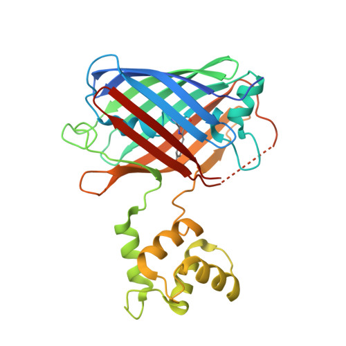
Fluorescent sensors for imaging of interstitial calcium.
Valiente-Gabioud, A.A., Garteizgogeascoa Suner, I., Idziak, A., Fabritius, A., Basquin, J., Angibaud, J., Nagerl, U.V., Singh, S.P., Griesbeck, O.(2023) Nat Commun 14: 6220-6220
- PubMed: 37798285 Search on PubMedSearch on PubMed Central
- DOI: https://doi.org/10.1038/s41467-023-41928-w
- Primary Citation Related Structures:
8C0T - PubMed Abstract:
Calcium in interstitial fluids is central to systemic physiology and a crucial ion pool for entry into cells through numerous plasma membrane channels. Its study has been limited by the scarcity of methods that allow monitoring in tight inter-cell spaces of living tissues. Here we present high performance ultra-low affinity genetically encoded calcium biosensors named GreenT-ECs. GreenT-ECs combine large fluorescence changes upon calcium binding and binding affinities (Kds) ranging from 0.8 mM to 2.9 mM, making them tuned to calcium concentrations in extracellular organismal fluids. We validated GreenT-ECs in rodent hippocampal neurons and transgenic zebrafish in vivo, where the sensors enabled monitoring homeostatic regulation of tissue interstitial calcium. GreenT-ECs may become useful for recording very large calcium transients and for imaging calcium homeostasis in inter-cell structures in live tissues and organisms.
- Max Planck Institute for Biological Intelligence, Tools for Bio-Imaging, Am Klopferspitz 18, 82152, Martinsried, Germany.
Organizational Affiliation: