Emerging variants of SARS-CoV-2 NSP10 highlight strong functional conservation of its binding to two non-structural proteins, NSP14 and NSP16.
Wang, H., Rizvi, S.R.A., Dong, D., Lou, J., Wang, Q., Sopipong, W., Su, Y., Najar, F., Agarwal, P.K., Kozielski, F., Haider, S.(2023) Elife 12
- PubMed: 38127066 Search on PubMedSearch on PubMed Central
- DOI: https://doi.org/10.7554/eLife.87884
- Primary Citation Related Structures:
8BZN - PubMed Abstract:
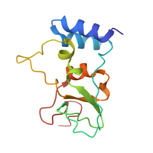
The coronavirus SARS-CoV-2 protects its RNA from being recognized by host immune responses by methylation of its 5' end, also known as capping. This process is carried out by two enzymes, non-structural protein 16 (NSP16) containing 2'-O-methyltransferase and NSP14 through its N7 methyltransferase activity, which are essential for the replication of the viral genome as well as evading the host's innate immunity. NSP10 acts as a crucial cofactor and stimulator of NSP14 and NSP16. To further understand the role of NSP10, we carried out a comprehensive analysis of >13 million globally collected whole-genome sequences (WGS) of SARS-CoV-2 obtained from the Global Initiative Sharing All Influenza Data (GISAID) and compared it with the reference genome Wuhan/WIV04/2019 to identify all currently known variants in NSP10. T12I, T102I, and A104V in NSP10 have been identified as the three most frequent variants and characterized using X-ray crystallography, biophysical assays, and enhanced sampling simulations. In contrast to other proteins such as spike and NSP6, NSP10 is significantly less prone to mutation due to its crucial role in replication. The functional effects of the variants were examined for their impact on the binding affinity and stability of both NSP14-NSP10 and NSP16-NSP10 complexes. These results highlight the limited changes induced by variant evolution in NSP10 and reflect on the critical roles NSP10 plays during the SARS-CoV-2 life cycle. These results also indicate that there is limited capacity for the virus to overcome inhibitors targeting NSP10 via the generation of variants in inhibitor binding pockets.
- Department of Pharmaceutical and Biological Chemistry, School of Pharmacy, University College London, London, United Kingdom.
Organizational Affiliation: