Download Mendeley
Structural insights into ligand recognition by the pleiotropic odorant-binding protein AgamOBP9.
Christodoulou, E., Stamati, E.C.V., Saitta, F., Papakyriakou, A., Fessas, D., Tsitsanou, K.E., Zographos, S.E.(2026) Int J Biol Macromol 372: 153025-153025
- PubMed: 42285453 Search on PubMed
- DOI: https://doi.org/10.1016/j.ijbiomac.2026.153025
- Primary Citation Related Structures:
29LI, 29LY, 29MC, 29MD, 8BY4 - PubMed Abstract:
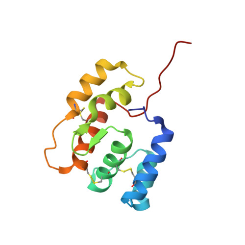
Odorant-binding proteins (OBPs) in mosquitoes play central roles in chemosensory perception and are attractive targets for vector control strategies. However, some OBPs are also expressed in non-olfactory tissues, suggesting a pleiotropic function that has not been sufficiently investigated. The crystal structures of the pleiotropic AgamOBP9 of the Anopheles gambiae mosquito were determined in complex with three plant-derived bioactive molecules: the phenylpropanoids n-butyl cinnamate and methyl eugenol (ME), as well as the monoterpene p-menthane-3, 8-diol (PMD). Fluorescence competitive binding assays and binding free-energy calculations identified that n-butyl cinnamate and ME bind with Ki values in the micromolar range, in contrast to the weak affinity observed for PMD. Structural analysis revealed an extended internal cavity comprising two distinct ligand-binding regions. One region, located at the bottom of the cavity, accommodated all investigated ligands without undergoing significant conformational changes, suggesting a structurally preordered binding pocket. The second region, situated at the cavity entrance, binds MPD and PEG, both used as crystallization agents, and may therefore serve as a recognition site for molecules with diverse chemical features. In the AgamOBP9-butyl cinnamate-MPD complex, MPD binding to the entrance-site induces rearrangements of surrounding residues, including Arg8, Arg15, Tyr32, Lys33, and Trp35. These interactions appear to promote the convergence of helices α1, α2, and the α2-α3 connecting loop toward a more "closed" protein conformation. The conformational flexibility of AgamOBP9 at the entrance region was further supported by differential scanning calorimetry and molecular dynamics simulations, which suggested the presence of two independent thermodynamic domains within the protein. Structural superposition with the homologous AaegOBP22-linoleic acid complex indicated that the AgamOBP9 cavity could accommodate fatty acids or other long-chain molecules spanning both binding regions, implying potential functions beyond conventional olfactory signaling. These findings provide new insights into the molecular basis of ligand recognition by AgamOBP9, which may guide the discovery of novel OBP9-targeting ligands.
- Institute of Chemical Biology, National Hellenic Research Foundation, 48 Vassileos Constantinou Avenue, 11635, Athens, Greece; Section of Pharmacognosy and Chemistry of Natural Products, Department of Pharmacy, National and Kapodistrian University of Athens, Athens, 15771, Greece.
Organizational Affiliation: