8BTY

Structure of the active form of ScpB, the C5a-peptidase from Streptococcus agalactiae.


Experimental Data Snapshot

  • Method: X-RAY DIFFRACTION
  • Resolution: 1.70 Å
  • R-Value Free: 0.216 
  • R-Value Work: 0.188 
  • R-Value Observed: 0.190 

wwPDB Validation   3D Report Full Report


This is version 1.1 of the entry. See complete history


Literature

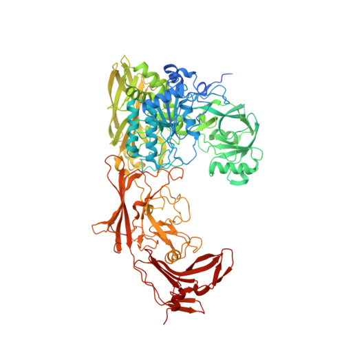
The 1.7 angstrom crystal structure of the C5a peptidase from Streptococcus agalactiae (ScpB) reveals an active site competent for catalysis.

Cullen, R.Tecza, M.Miclot, T.Behan, S.Jain, M.Avink, M.K.Cooney, J.C.Kagawa, T.F.

(2024) Proteins 92: 427-431

  • DOI: https://doi.org/10.1002/prot.26625
  • Primary Citation of Related Structures:  
    8BTY

  • PubMed Abstract: 

    A 1.7 Å structure is presented for an active form of the virulence factor ScpB, the C5a peptidase from Streptococcus agalactiae. The previously reported structure of the ScpB active site mutant exhibited a large separation (~20 Å) between the catalytic His and Ser residues. Significant differences are observed in the catalytic domain between the current and mutant ScpB structures resulting with a high RMSD (4.6 Å). The fold of the active form of ScpB is nearly identical to ScpA (RMSD 0.2 Å), the C5a-peptidase from Streptococcus pyogenes. Both ScpA and ScpB have comparable activity against human C5a, indicating neither enzyme require host proteins for C5a-ase activity. These studies are a first step in resolving reported differences in the specificities of these enzymes.


  • Organizational Affiliation

    Department of Biological Sciences, University of Limerick, Limerick, Ireland.


Macromolecules
Find similar proteins by:  (by identity cutoff)  |  3D Structure
Entity ID: 1
MoleculeChains Sequence LengthOrganismDetailsImage
C5a peptidase
A, B
1,001Streptococcus agalactiaeMutation(s): 0 
Gene Names: scpB
UniProt
Find proteins for Q53637 (Streptococcus agalactiae)
Explore Q53637 
Go to UniProtKB:  Q53637
Entity Groups  
Sequence Clusters30% Identity50% Identity70% Identity90% Identity95% Identity100% Identity
UniProt GroupQ53637
Sequence Annotations
Expand
  • Reference Sequence
Small Molecules
Ligands 5 Unique
IDChains Name / Formula / InChI Key2D Diagram3D Interactions
EPE
Query on EPE

Download Ideal Coordinates CCD File 
P [auth A]4-(2-HYDROXYETHYL)-1-PIPERAZINE ETHANESULFONIC ACID
C8 H18 N2 O4 S
JKMHFZQWWAIEOD-UHFFFAOYSA-N
MLA
Query on MLA

Download Ideal Coordinates CCD File 
C [auth A]MALONIC ACID
C3 H4 O4
OFOBLEOULBTSOW-UHFFFAOYSA-N
SO4
Query on SO4

Download Ideal Coordinates CCD File 
D [auth A]
G [auth A]
H [auth A]
I [auth A]
J [auth A]
D [auth A],
G [auth A],
H [auth A],
I [auth A],
J [auth A],
K [auth A],
L [auth A],
M [auth A],
N [auth A],
O [auth A],
Q [auth A]
SULFATE ION
O4 S
QAOWNCQODCNURD-UHFFFAOYSA-L
CA
Query on CA

Download Ideal Coordinates CCD File 
E [auth A]CALCIUM ION
Ca
BHPQYMZQTOCNFJ-UHFFFAOYSA-N
NA
Query on NA

Download Ideal Coordinates CCD File 
F [auth A]SODIUM ION
Na
FKNQFGJONOIPTF-UHFFFAOYSA-N
Experimental Data & Validation

Experimental Data

Unit Cell:
Length ( Å )Angle ( ˚ )
a = 167.152α = 90
b = 167.152β = 90
c = 141.485γ = 120
Software Package:
Software NamePurpose
PHENIXrefinement
XDSdata reduction
PHASERphasing
Cootmodel building

Structure Validation

View Full Validation Report


Entry History & Funding Information

Deposition Data


Funding OrganizationLocationGrant Number
Irish Research CouncilIrelandRS/2012/391
Science Foundation IrelandIrelandGrant 12/RC/2275_P2
Enterprise IrelandIrelandCF/2013/3336

Revision History  (Full details and data files)

  • Version 1.0: 2023-11-15
    Type: Initial release
  • Version 1.1: 2024-02-14
    Changes: Database references