


Crystal structure of Trichoplax Scribble PDZ2 domain
Maddumage, J.C., Humbert, P.O., Kvansakul, M.To be published.
Experimental Data Snapshot
Starting Model: experimental
View more details
wwPDB Validation 3D Report Full Report

Entity ID: 1 | |||||
|---|---|---|---|---|---|
| Molecule | Chains | Sequence Length | Organism | Details | Image |
| Leucine-rich repeat-containing protein 1 | 95 | Trichoplax sp. H2 | Mutation(s): 0 Gene Names: TrispH2_005790 | 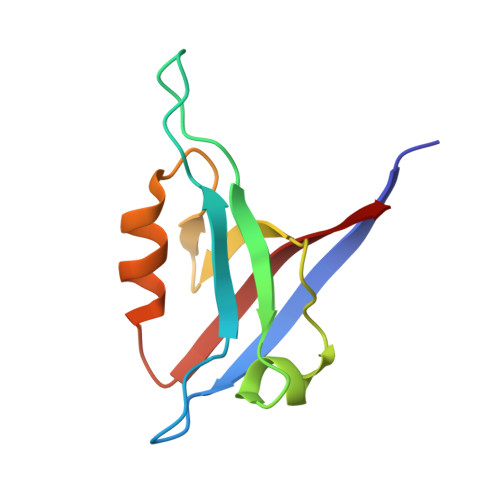 | |
| Length ( Å ) | Angle ( ˚ ) |
|---|---|
| a = 32.781 | α = 93.63 |
| b = 32.946 | β = 95.65 |
| c = 42.71 | γ = 113.74 |
| Software Name | Purpose |
|---|---|
| PHENIX | refinement |
| DIALS | data reduction |
| Aimless | data scaling |
| PHASER | phasing |
| Funding Organization | Location | Grant Number |
|---|---|---|
| National Health and Medical Research Council (NHMRC, Australia) | Australia | APP1103871 |
| Australian Research Council (ARC) | Australia | FT130101349 |


