8BF1

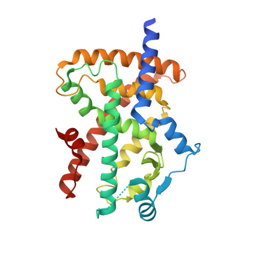
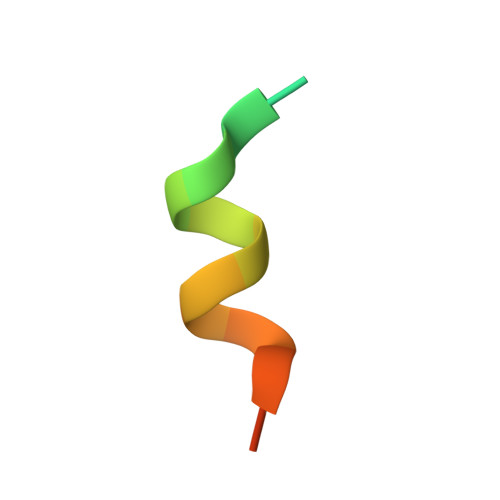
High-resolution structure of unliganded PPAR gamma in complex with the peptide PGC-1 alpha


Experimental Data Snapshot

  • Method: X-RAY DIFFRACTION
  • Resolution: 1.36 Å
  • R-Value Free: 0.181 
  • R-Value Work: 0.168 
  • R-Value Observed: 0.168 

Starting Model: experimental
View more details

wwPDB Validation   3D Report Full Report


This is version 1.1 of the entry. See complete history


Literature

Structural basis of the activation of PPAR gamma by the plasticizer metabolites MEHP and MINCH.

Useini, A.Engelberger, F.Kunze, G.Strater, N.

(2023) Environ Int 173: 107822-107822

  • DOI: https://doi.org/10.1016/j.envint.2023.107822
  • Primary Citation of Related Structures:  
    8BF1, 8BF2, 8BFF

  • PubMed Abstract: 

    Di-2-ethylhexyl phthalate (DEHP) and its substitute 1,2-cyclohexane dicarboxylic acid diisononyl ester (DINCH) are widely used as plasticizers but may have adverse health effects. Via hydrolysis of one of the two ester bonds in the human body, DEHP and DINCH form the monoesters MEHP and MINCH, respectively. Previous studies demonstrated binding of these metabolites to PPARγ and the induction of adipogenesis via this pathway. Detailed structural understanding of how these metabolites interact with PPARγ and thereby affect human health is lacking until now. We therefore characterized the binding modes of MINCH and MEHP to the ligand binding domain of PPARγ by X-ray crystallography and molecular dynamics (MD) simulations. Both compounds bind to the activating function-2 (AF-2) binding site via an interaction of the free carboxylates with the histidines 323 and 449, tyrosine 473 and serine 289. The alkyl chains form hydrophobic interactions with the tunnel next to cysteine 285. These binding modes are generally stable as demonstrated by the MD simulations and they resemble the complexation of fatty acids and their metabolites to the AF-2 site of PPARγ. Similar to the situation for these natural PPARγ agonists, the interaction of the free carboxylate groups of MEHP and MINCH with tyrosine 473 and surrounding residues stabilizes the AF-2 helix in the upward conformation. This state promotes binding of coactivator proteins and thus formation of the active complex for transcription of the specific target genes. Moreover, a comparison of the residues involved in binding of the plasticizer metabolites in vertebrate PPARγ orthologs shows that these compounds likely have similar effects in other species.


  • Organizational Affiliation

    Institute of Bioanalytical Chemistry, Centre for Biotechnology and Biomedicine, Leipzig University, Deutscher Platz 5, 04103 Leipzig, Germany.


Macromolecules
Find similar proteins by:  (by identity cutoff)  |  3D Structure
Entity ID: 1
MoleculeChains Sequence LengthOrganismDetailsImage
Peroxisome proliferator-activated receptor gamma283Homo sapiensMutation(s): 0 
Gene Names: PPARGNR1C3
UniProt & NIH Common Fund Data Resources
Find proteins for P37231 (Homo sapiens)
Explore P37231 
Go to UniProtKB:  P37231
PHAROS:  P37231
GTEx:  ENSG00000132170 
Entity Groups  
Sequence Clusters30% Identity50% Identity70% Identity90% Identity95% Identity100% Identity
UniProt GroupP37231
Sequence Annotations
Expand
  • Reference Sequence

Find similar proteins by:  Sequence   |   3D Structure  

Entity ID: 2
MoleculeChains Sequence LengthOrganismDetailsImage
Peroxisome proliferator-activated receptor gamma coactivator 1-alpha19Homo sapiensMutation(s): 0 
Gene Names: PPARGC1ALEM6PGC1PGC1APPARGC1
UniProt & NIH Common Fund Data Resources
Find proteins for Q9UBK2 (Homo sapiens)
Explore Q9UBK2 
Go to UniProtKB:  Q9UBK2
PHAROS:  Q9UBK2
GTEx:  ENSG00000109819 
Entity Groups  
Sequence Clusters30% Identity50% Identity70% Identity90% Identity95% Identity100% Identity
UniProt GroupQ9UBK2
Sequence Annotations
Expand
  • Reference Sequence
Experimental Data & Validation

Experimental Data

  • Method: X-RAY DIFFRACTION
  • Resolution: 1.36 Å
  • R-Value Free: 0.181 
  • R-Value Work: 0.168 
  • R-Value Observed: 0.168 
  • Space Group: P 1 21 1
Unit Cell:
Length ( Å )Angle ( ˚ )
a = 43.706α = 90
b = 54.209β = 106.73
c = 66.089γ = 90
Software Package:
Software NamePurpose
PHENIXrefinement
Aimlessdata scaling
PDB_EXTRACTdata extraction
XDSdata reduction
Cootmodel building

Structure Validation

View Full Validation Report


Entry History & Funding Information

Deposition Data


Funding OrganizationLocationGrant Number
German Research Foundation (DFG)GermanySfb1052-Z6

Revision History  (Full details and data files)

  • Version 1.0: 2023-03-08
    Type: Initial release
  • Version 1.1: 2024-02-07
    Changes: Data collection, Refinement description