8B5K

Structure of haloalkane dehalogenase DmmarA from Mycobacterium marinum at pH 6.5


Experimental Data Snapshot

  • Method: X-RAY DIFFRACTION
  • Resolution: 1.85 Å
  • R-Value Free: 0.241 
  • R-Value Work: 0.191 
  • R-Value Observed: 0.194 

wwPDB Validation   3D Report Full Report


Ligand Structure Quality Assessment 


This is version 1.1 of the entry. See complete history


Literature

Atypical homodimerization revealed by the structure of the (S)-enantioselective haloalkane dehalogenase DmmarA from Mycobacterium marinum.

Snajdarova, K.Marques, S.M.Damborsky, J.Bednar, D.Marek, M.

(2023) Acta Crystallogr D Struct Biol 79: 956-970

  • DOI: https://doi.org/10.1107/S2059798323006642
  • Primary Citation of Related Structures:  
    8B5K, 8B5O

  • PubMed Abstract: 

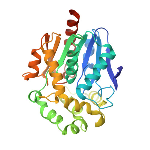
    Haloalkane dehalogenases (HLDs) are a family of α/β-hydrolase fold enzymes that employ S N 2 nucleophilic substitution to cleave the carbon-halogen bond in diverse chemical structures, the biological role of which is still poorly understood. Atomic-level knowledge of both the inner organization and supramolecular complexation of HLDs is thus crucial to understand their catalytic and noncatalytic functions. Here, crystallographic structures of the (S)-enantioselective haloalkane dehalogenase DmmarA from the waterborne pathogenic microbe Mycobacterium marinum were determined at 1.6 and 1.85 Å resolution. The structures show a canonical αβα-sandwich HLD fold with several unusual structural features. Mechanistically, the atypical composition of the proton-relay catalytic triad (aspartate-histidine-aspartate) and uncommon active-site pocket reveal the molecular specificities of a catalytic apparatus that exhibits a rare (S)-enantiopreference. Additionally, the structures reveal a previously unobserved mode of symmetric homodimerization, which is predominantly mediated through unusual L5-to-L5 loop interactions. This homodimeric association in solution is confirmed experimentally by data obtained from small-angle X-ray scattering. Utilizing the newly determined structures of DmmarA, molecular modelling techniques were employed to elucidate the underlying mechanism behind its uncommon enantioselectivity. The (S)-preference can be attributed to the presence of a distinct binding pocket and variance in the activation barrier for nucleophilic substitution.


  • Organizational Affiliation

    Loschmidt Laboratories, Department of Experimental Biology and RECETOX, Faculty of Science, Masaryk University, Kamenice 5, Building A13, 625 00 Brno, Czech Republic.


Macromolecules
Find similar proteins by:  (by identity cutoff)  |  3D Structure
Entity ID: 1
MoleculeChains Sequence LengthOrganismDetailsImage
Haloalkane dehalogenase DhaA
A, B, C, D
296Mycobacterium marinumMutation(s): 0 
Gene Names: dhaAMMAR_4113
UniProt
Find proteins for B2HR89 (Mycobacterium marinum (strain ATCC BAA-535 / M))
Explore B2HR89 
Go to UniProtKB:  B2HR89
Entity Groups  
Sequence Clusters30% Identity50% Identity70% Identity90% Identity95% Identity100% Identity
UniProt GroupB2HR89
Sequence Annotations
Expand
  • Reference Sequence
Small Molecules
Ligands 4 Unique
IDChains Name / Formula / InChI Key2D Diagram3D Interactions
B3P (Subject of Investigation/LOI)
Query on B3P

Download Ideal Coordinates CCD File 
V [auth D]2-[3-(2-HYDROXY-1,1-DIHYDROXYMETHYL-ETHYLAMINO)-PROPYLAMINO]-2-HYDROXYMETHYL-PROPANE-1,3-DIOL
C11 H26 N2 O6
HHKZCCWKTZRCCL-UHFFFAOYSA-N
TRS (Subject of Investigation/LOI)
Query on TRS

Download Ideal Coordinates CCD File 
R [auth C]2-AMINO-2-HYDROXYMETHYL-PROPANE-1,3-DIOL
C4 H12 N O3
LENZDBCJOHFCAS-UHFFFAOYSA-O
GOL (Subject of Investigation/LOI)
Query on GOL

Download Ideal Coordinates CCD File 
E [auth A]
F [auth A]
H [auth A]
I [auth B]
J [auth B]
E [auth A],
F [auth A],
H [auth A],
I [auth B],
J [auth B],
M [auth C],
P [auth C],
Q [auth C],
S [auth C]
GLYCEROL
C3 H8 O3
PEDCQBHIVMGVHV-UHFFFAOYSA-N
FMT (Subject of Investigation/LOI)
Query on FMT

Download Ideal Coordinates CCD File 
G [auth A]
K [auth B]
L [auth C]
N [auth C]
O [auth C]
G [auth A],
K [auth B],
L [auth C],
N [auth C],
O [auth C],
T [auth D],
U [auth D]
FORMIC ACID
C H2 O2
BDAGIHXWWSANSR-UHFFFAOYSA-N
Experimental Data & Validation

Experimental Data

  • Method: X-RAY DIFFRACTION
  • Resolution: 1.85 Å
  • R-Value Free: 0.241 
  • R-Value Work: 0.191 
  • R-Value Observed: 0.194 
  • Space Group: P 1 21 1
Unit Cell:
Length ( Å )Angle ( ˚ )
a = 91.816α = 90
b = 61.381β = 106.26
c = 106.689γ = 90
Software Package:
Software NamePurpose
XDSdata reduction
Aimlessdata scaling
PHASERphasing
PHENIXrefinement
PDB_EXTRACTdata extraction

Structure Validation

View Full Validation Report


Ligand Structure Quality Assessment 


Entry History & Funding Information

Deposition Data


Funding OrganizationLocationGrant Number
Czech Science FoundationCzech Republic22-09853S

Revision History  (Full details and data files)

  • Version 1.0: 2023-08-30
    Type: Initial release
  • Version 1.1: 2024-03-13
    Changes: Database references