A DNA damage-induced phosphorylation circuit enhances Mec1 ATR Ddc2 ATRIP recruitment to Replication Protein A.
Yates, L.A., Tannous, E.A., Morgan, R.M., Burgers, P.M., Zhang, X.(2023) Proc Natl Acad Sci U S A 120: e2300150120-e2300150120
- PubMed: 36996117 Search on PubMedSearch on PubMed Central
- DOI: https://doi.org/10.1073/pnas.2300150120
- Primary Citation Related Structures:
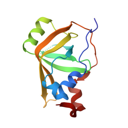
8B4J, 8B4K - PubMed Abstract:
The cell cycle checkpoint kinase Mec1 ATR and its integral partner Ddc2 ATRIP are vital for the DNA damage and replication stress response. Mec1-Ddc2 "senses" single-stranded DNA (ssDNA) by being recruited to the ssDNA binding Replication Protein A (RPA) via Ddc2. In this study, we show that a DNA damage-induced phosphorylation circuit modulates checkpoint recruitment and function. We demonstrate that Ddc2-RPA interactions modulate the association between RPA and ssDNA and that Rfa1-phosphorylation aids in the further recruitment of Mec1-Ddc2. We also uncover an underappreciated role for Ddc2 phosphorylation that enhances its recruitment to RPA-ssDNA that is important for the DNA damage checkpoint in yeast. The crystal structure of a phosphorylated Ddc2 peptide in complex with its RPA interaction domain provides molecular details of how checkpoint recruitment is enhanced, which involves Zn 2+ . Using electron microscopy and structural modeling approaches, we propose that Mec1-Ddc2 complexes can form higher order assemblies with RPA when Ddc2 is phosphorylated. Together, our results provide insight into Mec1 recruitment and suggest that formation of supramolecular complexes of RPA and Mec1-Ddc2, modulated by phosphorylation, would allow for rapid clustering of damage foci to promote checkpoint signaling.
- Section of Structural Biology, Department of Infectious Disease, Imperial College London, South Kensington, London SW7 2AZ, United Kingdom.
Organizational Affiliation: