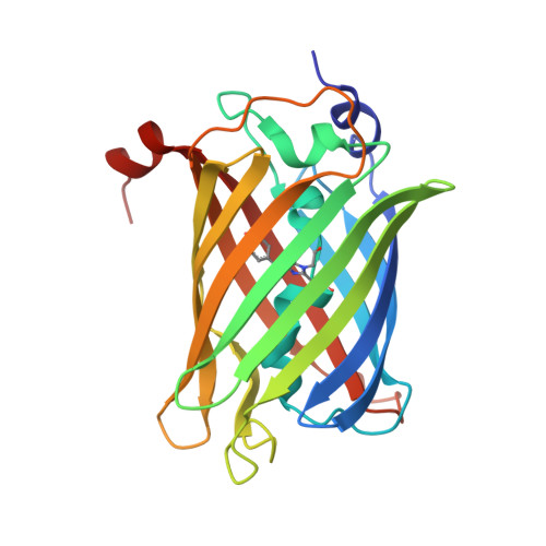
Structure of the weakly red fluorescent protein csiFP4 from Clytia simplex
Depernet, H., Engilberge, S., Lambert, G., Gotthard, G., Shaner, N., Royant, A.To be published.
Experimental Data Snapshot
Starting Model: experimental
View more details
wwPDB Validation 3D Report Full Report

| Ligands 2 Unique | |||||
|---|---|---|---|---|---|
| ID | Chains | Name / Formula / InChI Key | 2D Diagram | 3D Interactions | |
| SO4 Download:Ideal Coordinates CCD File | AA [auth C] BA [auth C] E [auth A] EA [auth D] F [auth A] | SULFATE ION O4 S QAOWNCQODCNURD-UHFFFAOYSA-L |  | ||
| CL Download:Ideal Coordinates CCD File | CA [auth C] DA [auth C] HA [auth D] IA [auth D] N [auth A] | CHLORIDE ION Cl VEXZGXHMUGYJMC-UHFFFAOYSA-M |  | ||
| Modified Residues 1 Unique | |||||
|---|---|---|---|---|---|
| ID | Chains | Type | Formula | 2D Diagram | Parent |
| CRQ Query on CRQ | A, B, C, D | L-PEPTIDE LINKING | C16 H16 N4 O5 |  | GLN, TYR, GLY |
| Length ( Å ) | Angle ( ˚ ) |
|---|---|
| a = 182.904 | α = 90 |
| b = 182.904 | β = 90 |
| c = 155.87 | γ = 120 |
| Software Name | Purpose |
|---|---|
| PHENIX | refinement |
| PDB_EXTRACT | data extraction |
| XDS | data reduction |
| Aimless | data scaling |
| PHASER | phasing |
| Funding Organization | Location | Grant Number |
|---|---|---|
| Not funded | -- |