8AS9

Crystal structure of the talin-KANK1 complex


Experimental Data Snapshot

  • Method: X-RAY DIFFRACTION
  • Resolution: 3.40 Å
  • R-Value Free: 0.285 
  • R-Value Work: 0.245 
  • R-Value Observed: 0.247 

wwPDB Validation   3D Report Full Report


This is version 1.3 of the entry. See complete history


Literature

The structural basis of the talin-KANK1 interaction that coordinates the actin and microtubule cytoskeletons at focal adhesions.

Li, X.Goult, B.T.Ballestrem, C.Zacharchenko, T.

(2023) Open Biol 13: 230058-230058

  • DOI: https://doi.org/10.1098/rsob.230058
  • Primary Citation of Related Structures:  
    8AS9

  • PubMed Abstract: 

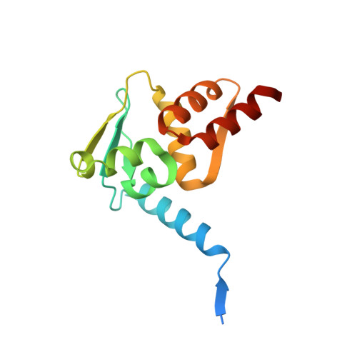
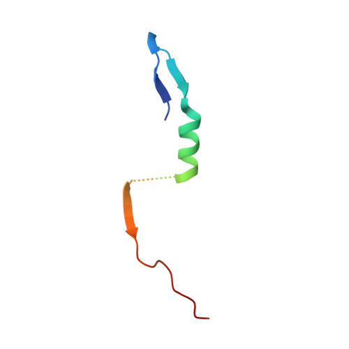
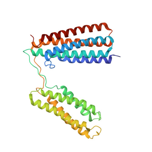
    Adhesion between cells and the extracellular matrix is mediated by heterodimeric ( αβ ) integrin receptors that are intracellularly linked to the contractile actomyosin machinery. One of the proteins that control this link is talin, which organizes cytosolic signalling proteins into discrete complexes on β-integrin tails referred to as focal adhesions (FAs). The adapter protein KANK1 binds to talin in the region of FAs known as the adhesion belt. Here, we adapted a non-covalent crystallographic chaperone to resolve the talin-KANK1 complex. This structure revealed that the talin binding KN region of KANK1 contains a novel motif where a β-hairpin stabilizes the α-helical region, explaining both its specific interaction with talin R7 and high affinity. Single point mutants in KANK1 identified from the structure abolished the interaction and enabled us to examine KANK1 enrichment in the adhesion belt. Strikingly, in cells expressing a constitutively active form of vinculin that keeps the FA structure intact even in the presence of myosin inhibitors, KANK1 localizes throughout the entire FA structure even when actomyosin tension is released. We propose a model whereby actomyosin forces on talin eliminate KANK1 from talin binding in the centre of FAs while retaining it at the adhesion periphery.


  • Organizational Affiliation

    Wellcome Centre for Cell-Matrix Research, Faculty of Biology, Medicine and Health, University of Manchester, Dover Street, Manchester M13 9PT, UK.


Macromolecules
Find similar proteins by:  (by identity cutoff)  |  3D Structure
Entity ID: 1
MoleculeChains Sequence LengthOrganismDetailsImage
B-cell lymphoma 6 protein
A, B
137Homo sapiensMutation(s): 3 
Gene Names: BCL6BCL5LAZ3ZBTB27ZNF51
UniProt & NIH Common Fund Data Resources
Find proteins for P41182 (Homo sapiens)
Explore P41182 
Go to UniProtKB:  P41182
PHAROS:  P41182
GTEx:  ENSG00000113916 
Entity Groups  
Sequence Clusters30% Identity50% Identity70% Identity90% Identity95% Identity100% Identity
UniProt GroupP41182
Sequence Annotations
Expand
  • Reference Sequence
Find similar proteins by:  (by identity cutoff)  |  3D Structure
Entity ID: 2
MoleculeChains Sequence LengthOrganismDetailsImage
KN-motif NCoR1 BBD fusion,Nuclear receptor corepressor 1C [auth D]51Homo sapiensMutation(s): 0 
UniProt & NIH Common Fund Data Resources
Find proteins for Q14678 (Homo sapiens)
Explore Q14678 
Go to UniProtKB:  Q14678
PHAROS:  Q14678
GTEx:  ENSG00000107104 
Find proteins for O75376 (Homo sapiens)
Explore O75376 
Go to UniProtKB:  O75376
PHAROS:  O75376
GTEx:  ENSG00000141027 
Entity Groups  
Sequence Clusters30% Identity50% Identity70% Identity90% Identity95% Identity100% Identity
UniProt GroupsO75376Q14678
Sequence Annotations
Expand
  • Reference Sequence
Find similar proteins by:  (by identity cutoff)  |  3D Structure
Entity ID: 3
MoleculeChains Sequence LengthOrganismDetailsImage
Talin-1D [auth C]309Mus musculusMutation(s): 0 
Gene Names: Tln1Tln
UniProt
Find proteins for P26039 (Mus musculus)
Explore P26039 
Go to UniProtKB:  P26039
Entity Groups  
Sequence Clusters30% Identity50% Identity70% Identity90% Identity95% Identity100% Identity
UniProt GroupP26039
Sequence Annotations
Expand
  • Reference Sequence
Experimental Data & Validation

Experimental Data

  • Method: X-RAY DIFFRACTION
  • Resolution: 3.40 Å
  • R-Value Free: 0.285 
  • R-Value Work: 0.245 
  • R-Value Observed: 0.247 
  • Space Group: H 3 2
Unit Cell:
Length ( Å )Angle ( ˚ )
a = 207.022α = 90
b = 207.022β = 90
c = 151.859γ = 120
Software Package:
Software NamePurpose
PHENIXrefinement
XDSdata reduction
SCALAdata scaling
PHASERphasing

Structure Validation

View Full Validation Report


Entry History & Funding Information

Deposition Data


Funding OrganizationLocationGrant Number
Not fundedUnited Kingdom--

Revision History  (Full details and data files)

  • Version 1.0: 2023-06-07
    Type: Initial release
  • Version 1.1: 2023-07-12
    Changes: Database references
  • Version 1.2: 2023-11-15
    Changes: Data collection, Source and taxonomy
  • Version 1.3: 2024-02-07
    Changes: Refinement description