Crystal Structures of the Clostridium botulinum Neurotoxin A6 Cell Binding Domain Alone and in Complex with GD1a Reveal Significant Conformational Flexibility.
Gregory, K.S., Newell, A.R., Mojanaga, O.O., Liu, S.M., Acharya, K.R.(2022) Int J Mol Sci 23
- PubMed: 36077016 Search on PubMedSearch on PubMed Central
- DOI: https://doi.org/10.3390/ijms23179620
- Primary Citation Related Structures:
8AGK, 8ALP - PubMed Abstract:
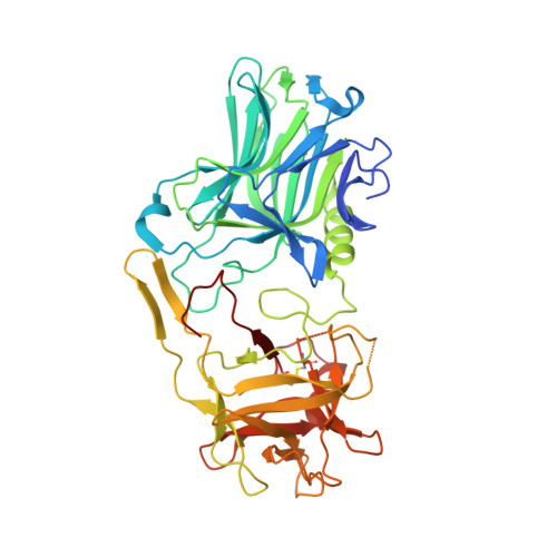
Clostridium botulinum neurotoxin A (BoNT/A) targets the soluble N -ethylmaleimide-sensitive factor attachment protein receptor (SNARE) complex, by cleaving synaptosomal-associated protein of 25 kDa size ( SNAP - 25 ). Cleavage of SNAP-25 results in flaccid paralysis due to repression of synaptic transmission at the neuromuscular junction. This activity has been exploited to treat a range of diseases associated with hypersecretion of neurotransmitters, with formulations of BoNT/A commercially available as therapeutics. Generally, BoNT activity is facilitated by three essential domains within the molecule, the cell binding domain (H C ), the translocation domain (H N ), and the catalytic domain (LC). The H C, which consists of an N-terminal (H CN ) and a C-terminal (H CC ) subdomain, is responsible for BoNT's high target specificity where it forms a dual-receptor complex with synaptic vesicle protein 2 (SV2) and a ganglioside receptor on the surface of motor neurons. In this study, we have determined the crystal structure of botulinum neurotoxin A6 cell binding domain (H C /A6) in complex with GD1a and describe the interactions involved in ganglioside binding. We also present a new crystal form of wild type H C /A6 (crystal form II) where a large 'hinge motion' between the H CN and H CC subdomains is observed. These structures, along with a comparison to the previously determined wild type crystal structure of H C /A6 (crystal form I), reveals the degree of conformational flexibility exhibited by H C /A6.
- Department of Life Sciences, University of Bath, Claverton Down, Bath BA2 7AY, UK.
Organizational Affiliation: