8AG0

Crystal structure of mutant PRELID3a-TRIAP1 complex - R53E


Experimental Data Snapshot

  • Method: X-RAY DIFFRACTION
  • Resolution: 2.70 Å
  • R-Value Free: 0.304 
  • R-Value Work: 0.213 
  • R-Value Observed: 0.218 

wwPDB Validation   3D Report Full Report


This is version 1.1 of the entry. See complete history


Literature

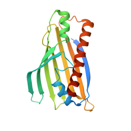
An intermolecular hydrogen bonded network in the PRELID-TRIAP protein family plays a role in lipid sensing.

Miliara, X.Tatsuta, T.Eiyama, A.Langer, T.Rouse, S.L.Matthews, S.

(2022) Biochim Biophys Acta Proteins Proteom 1871: 140867-140867

  • DOI: https://doi.org/10.1016/j.bbapap.2022.140867
  • Primary Citation of Related Structures:  
    8AG0

  • PubMed Abstract: 

    The PRELID-TRIAP1 family of proteins is responsible for lipid transfer in mitochondria. Multiple structures have been resolved of apo and lipid substrate bound forms, allowing us to begin to piece together the molecular level details of the full lipid transfer cycle. Here, we used molecular dynamics simulations to demonstrate that the lipid binding is mediated by an extended, water-mediated hydrogen bonding network. A key mutation, R53E, was found to disrupt this network, causing lipid to be released from the complex. The X-ray crystal structure of R53E was captured in a fully closed and apo state. Lipid transfer assays and molecular simulations allow us to interpret the observed conformation in the context of the biological role. Together, our work provides further understanding of the mechanistic control of lipid transport by PRELID-TRIAP1 in mitochondria.


  • Organizational Affiliation

    Department of Life Sciences, Imperial College London, South Kensington, London SW7 2AZ, UK.


Macromolecules
Find similar proteins by:  (by identity cutoff)  |  3D Structure
Entity ID: 1
MoleculeChains Sequence LengthOrganismDetailsImage
PRELI domain containing protein 3A186Homo sapiensMutation(s): 1 
Gene Names: PRELID3AC18orf43SLMO1
UniProt & NIH Common Fund Data Resources
Find proteins for Q96N28 (Homo sapiens)
Explore Q96N28 
Go to UniProtKB:  Q96N28
PHAROS:  Q96N28
GTEx:  ENSG00000141391 
Entity Groups  
Sequence Clusters30% Identity50% Identity70% Identity90% Identity95% Identity100% Identity
UniProt GroupQ96N28
Sequence Annotations
Expand
  • Reference Sequence
Find similar proteins by:  (by identity cutoff)  |  3D Structure
Entity ID: 2
MoleculeChains Sequence LengthOrganismDetailsImage
Maltose/maltodextrin-binding periplasmic protein,TP53-regulated inhibitor of apoptosis 1446Homo sapiensMutation(s): 0 
Gene Names: malEb4034JW3994TRIAP115E1.1HSPC132
UniProt & NIH Common Fund Data Resources
Find proteins for P0AEX9 (Escherichia coli (strain K12))
Explore P0AEX9 
Go to UniProtKB:  P0AEX9
Find proteins for O43715 (Homo sapiens)
Explore O43715 
Go to UniProtKB:  O43715
PHAROS:  O43715
GTEx:  ENSG00000170855 
Entity Groups  
Sequence Clusters30% Identity50% Identity70% Identity90% Identity95% Identity100% Identity
UniProt GroupsO43715P0AEX9
Sequence Annotations
Expand
  • Reference Sequence
Oligosaccharides

Help

Entity ID: 3
MoleculeChains Length2D Diagram Glycosylation3D Interactions
alpha-D-glucopyranose-(1-4)-beta-D-glucopyranoseC [auth D]2N/A
Glycosylation Resources
GlyTouCan:  G66120GK
GlyCosmos:  G66120GK
Experimental Data & Validation

Experimental Data

  • Method: X-RAY DIFFRACTION
  • Resolution: 2.70 Å
  • R-Value Free: 0.304 
  • R-Value Work: 0.213 
  • R-Value Observed: 0.218 
  • Space Group: P 1 21 1
Unit Cell:
Length ( Å )Angle ( ˚ )
a = 62.3α = 90
b = 66.815β = 96.46
c = 82.14γ = 90
Software Package:
Software NamePurpose
PHENIXrefinement
MOSFLMdata reduction
SCALAdata scaling
PHASERphasing

Structure Validation

View Full Validation Report


Entry History & Funding Information

Deposition Data


Funding OrganizationLocationGrant Number
Medical Research Council (MRC, United Kingdom)United KingdomMR/M019403/1

Revision History  (Full details and data files)

  • Version 1.0: 2022-11-23
    Type: Initial release
  • Version 1.1: 2024-01-31
    Changes: Data collection, Refinement description