8AA6 | pdb_00008aa6

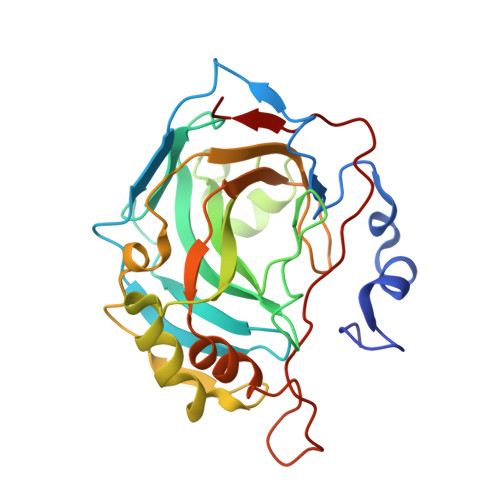
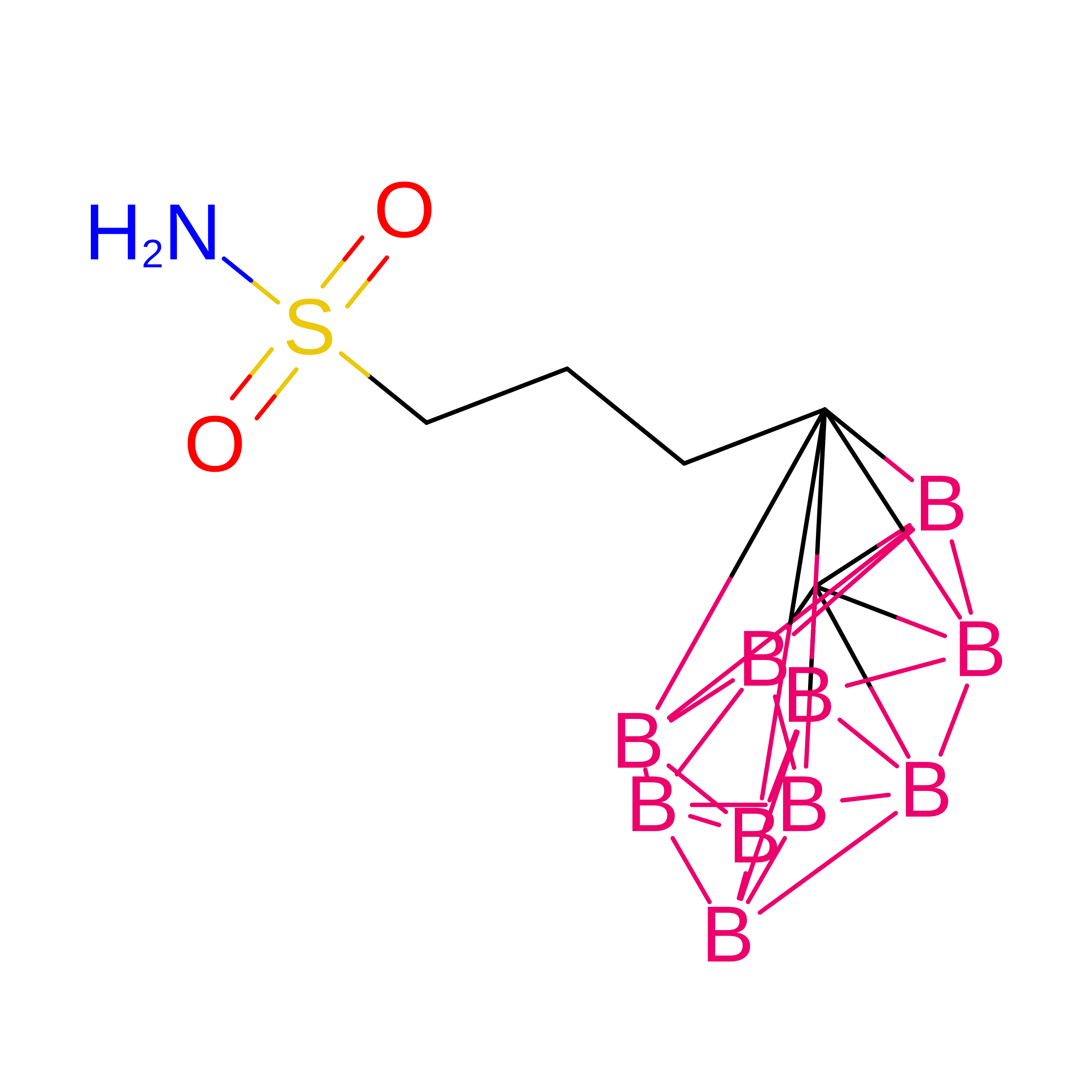
CAII in complex with meta-carboran-propylsulfonamid


Experimental Data Snapshot

  • Method: X-RAY DIFFRACTION
  • Resolution: 1.15 Å
  • R-Value Free: 
    0.180 (Depositor), 0.174 (DCC) 
  • R-Value Work: 
    0.162 (Depositor), 0.160 (DCC) 
  • R-Value Observed: 
    0.162 (Depositor) 

Starting Model: experimental
View more details

wwPDB Validation 3D Report Full Report

Validation slider image for 8AA6

Ligand Structure Quality Assessment 


This is version 1.1 of the entry. See complete history. 

Literature

B-H⋯ pi and C-H⋯ pi interactions in protein-ligand complexes: carbonic anhydrase II inhibition by carborane sulfonamides.

Fanfrlik, J., Brynda, J., Kugler, M., Lepsik, M., Pospisilova, K., Holub, J., Hnyk, D., Nekvinda, J., Gruner, B., Rezacova, P.

(2023) Phys Chem Chem Phys 25: 1728-1733

  • DOI: https://doi.org/10.1039/d2cp04673c
  • Primary Citation Related Structures: 
    8AA6, 8AAE

  • PubMed Abstract: 

    Among non-covalent interactions, B-H⋯π and C-H⋯π hydrogen bonding is rather weak and less studied. Nevertheless, since both can affect the energetics of protein-ligand binding, their understanding is an important prerequisite for reliable predictions of affinities. Through a combination of high-resolution X-ray crystallography and quantum-chemical calculations on carbonic anhydrase II/carborane-based inhibitor systems, this paper provides the first example of B-H⋯π hydrogen bonding in a protein-ligand complex. It shows that the B-H⋯π interaction is stabilized by dispersion, followed by electrostatics. Furthermore, it demonstrates that the similar C-H⋯π interaction is twice as strong, with a slightly smaller contribution of dispersion and a slightly higher contribution of electrostatics. Such a detailed insight will facilitate the rational design of future protein ligands, controlling these types of non-covalent interactions.


  • Organizational Affiliation: 
    • Institute of Organic Chemistry and Biochemistry of the Czech Academy of Sciences, Flemingovo nam. 2, 166 10, Prague 6, Czech Republic. fanfrlik@uochb.cas.cz.

Macromolecule Content 

  • Total Structure Weight: 29.61 kDa 
  • Atom Count: 2,360 
  • Modeled Residue Count: 257 
  • Deposited Residue Count: 260 
  • Unique protein chains: 1

Macromolecules

Find similar proteins by:|  3D Structure
Entity ID: 1
MoleculeChains  Sequence LengthOrganismDetailsImage
Carbonic anhydrase 2260Homo sapiensMutation(s): 0 
Gene Names: CA2
EC: 4.2.1.1 (PDB Primary Data), 4.2.1.69 (UniProt)
UniProt & NIH Common Fund Data Resources
Find proteins for P00918 (Homo sapiens)
Explore P00918 
Go to UniProtKB:  P00918
PHAROS:  P00918
GTEx:  ENSG00000104267 
Entity Groups
Sequence Clusters30% Identity50% Identity70% Identity90% Identity95% Identity100% Identity
UniProt GroupP00918
Sequence Annotations
Expand
Reference Sequence

Experimental Data & Validation

Experimental Data

  • Method: X-RAY DIFFRACTION
  • Resolution: 1.15 Å
  • R-Value Free:  0.180 (Depositor), 0.174 (DCC) 
  • R-Value Work:  0.162 (Depositor), 0.160 (DCC) 
  • R-Value Observed: 0.162 (Depositor) 
Space Group: P 1 21 1
Unit Cell:
Length ( Å )Angle ( ˚ )
a = 42.172α = 90
b = 41.432β = 104.33
c = 72.165γ = 90
Software Package:
Software NamePurpose
XDSdata reduction
XSCALEdata scaling
REFMACrefinement
PDB_EXTRACTdata extraction
MOLREPphasing

Structure Validation

View Full Validation Report


Ligand Structure Quality Assessment 


Entry History 

& Funding Information

Deposition Data


Funding OrganizationLocationGrant Number
Technology Agency of the Czech RepublicCzech RepublicTE01020028
Ministry of Education, Youth and Sports of the Czech RepublicCzech RepublicCZ.02.1.01/0.0/0.0/16_019/0000868

Revision History  (Full details and data files)

  • Version 1.0: 2023-05-10
    Type: Initial release
  • Version 1.1: 2024-02-07
    Changes: Data collection, Refinement description