X-ray structure solution of amaryllis lectin by molecular replacement with only 4% of the total diffracting matter.
Chantalat, L., Wood, S.D., Rizkallah, P., Reynolds, C.D.(1996) Acta Crystallogr D Biol Crystallogr 52: 1146-1152
- PubMed: 15299575 Search on PubMed
- DOI: https://doi.org/10.1107/S090744499600546X
- Primary Citation Related Structures:
8A9M - PubMed Abstract:
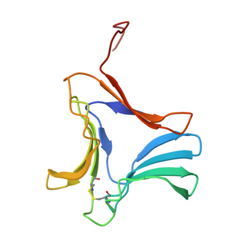
It is often the case that analogous proteins from different species crystallize in a different form. These structures can usually be easily solved by the molecular-replacement (MR) technique, as the protein folding is very often conserved. However, the results from MR become more uncertain as the proportion of diffracting matter decreases as a result of multimericity and/or absence of some of the atoms in the model. In this paper results are presented on the structure solution of amaryllis lectin (109 residues per monomer) containing two protein molecules in the asymmetric unit. The structure was solved by MR using the Calpha coordinates of one monomer from snowdrop lectin which has 85% amino-acid sequence identity to amaryllis lectin. This represents only 6% of the non-H atoms of the protein molecule to be used for structure determination and it is a major improvement on previous reports. Further calculations were carried out in order to establish the minimum number of atoms which could be included in the model before a clear solution to the MR problem was revealed. This study showed that the structure of amaryllis lectin could still have been solved easily with 3.85% of the model, which even in the most favourable cases, will probably constitute a minimum for molecular-replacement structure solution.
- Biophysics Department, School of Biomolecular Sciences, Liverpool John Moores University, England.
Organizational Affiliation: