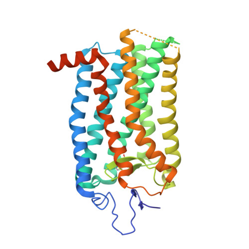
8A6C

1 picosecond light activated crystal structure of bovine rhodopsin in Lipidic Cubic Phase


Experimental Data Snapshot

  • Method: X-RAY DIFFRACTION
  • Resolution: 1.80 Å
  • R-Value Free: 0.398 
  • R-Value Work: 0.350 
  • R-Value Observed: 0.351 

wwPDB Validation   3D Report Full Report


Ligand Structure Quality Assessment 


This is version 1.3 of the entry. See complete history


Literature

Ultrafast structural changes direct the first molecular events of vision.

Gruhl, T.Weinert, T.Rodrigues, M.J.Milne, C.J.Ortolani, G.Nass, K.Nango, E.Sen, S.Johnson, P.J.M.Cirelli, C.Furrer, A.Mous, S.Skopintsev, P.James, D.Dworkowski, F.Bath, P.Kekilli, D.Ozerov, D.Tanaka, R.Glover, H.Bacellar, C.Brunle, S.Casadei, C.M.Diethelm, A.D.Gashi, D.Gotthard, G.Guixa-Gonzalez, R.Joti, Y.Kabanova, V.Knopp, G.Lesca, E.Ma, P.Martiel, I.Muhle, J.Owada, S.Pamula, F.Sarabi, D.Tejero, O.Tsai, C.J.Varma, N.Wach, A.Boutet, S.Tono, K.Nogly, P.Deupi, X.Iwata, S.Neutze, R.Standfuss, J.Schertler, G.Panneels, V.

(2023) Nature 615: 939-944

  • DOI: https://doi.org/10.1038/s41586-023-05863-6
  • Primary Citation of Related Structures:  
    7ZBC, 7ZBE, 8A6C, 8A6D, 8A6E

  • PubMed Abstract: 

    Vision is initiated by the rhodopsin family of light-sensitive G protein-coupled receptors (GPCRs) 1 . A photon is absorbed by the 11-cis retinal chromophore of rhodopsin, which isomerizes within 200 femtoseconds to the all-trans conformation 2 , thereby initiating the cellular signal transduction processes that ultimately lead to vision. However, the intramolecular mechanism by which the photoactivated retinal induces the activation events inside rhodopsin remains experimentally unclear. Here we use ultrafast time-resolved crystallography at room temperature 3 to determine how an isomerized twisted all-trans retinal stores the photon energy that is required to initiate the protein conformational changes associated with the formation of the G protein-binding signalling state. The distorted retinal at a 1-ps time delay after photoactivation has pulled away from half of its numerous interactions with its binding pocket, and the excess of the photon energy is released through an anisotropic protein breathing motion in the direction of the extracellular space. Notably, the very early structural motions in the protein side chains of rhodopsin appear in regions that are involved in later stages of the conserved class A GPCR activation mechanism. Our study sheds light on the earliest stages of vision in vertebrates and points to fundamental aspects of the molecular mechanisms of agonist-mediated GPCR activation.


  • Organizational Affiliation

    Division of Biology and Chemistry, Laboratory for Biomolecular Research, Paul Scherrer Institute, Villigen PSI, Switzerland.


Macromolecules
Find similar proteins by:  (by identity cutoff)  |  3D Structure
Entity ID: 1
MoleculeChains Sequence LengthOrganismDetailsImage
Rhodopsin
A, B
348Bos taurusMutation(s): 0 
Membrane Entity: Yes 
UniProt
Find proteins for P02699 (Bos taurus)
Explore P02699 
Go to UniProtKB:  P02699
Entity Groups  
Sequence Clusters30% Identity50% Identity70% Identity90% Identity95% Identity100% Identity
UniProt GroupP02699
Sequence Annotations
Expand
  • Reference Sequence
Oligosaccharides

Help

Entity ID: 2
MoleculeChains Length2D Diagram Glycosylation3D Interactions
2-acetamido-2-deoxy-beta-D-glucopyranose-(1-4)-2-acetamido-2-deoxy-beta-D-glucopyranose
C, D
2N-Glycosylation
Glycosylation Resources
GlyTouCan:  G42666HT
GlyCosmos:  G42666HT
GlyGen:  G42666HT
Small Molecules
Ligands 6 Unique
IDChains Name / Formula / InChI Key2D Diagram3D Interactions
OLC
Query on OLC

Download Ideal Coordinates CCD File 
I [auth A]
J [auth A]
K [auth A]
L [auth A]
M [auth A]
I [auth A],
J [auth A],
K [auth A],
L [auth A],
M [auth A],
O [auth A],
P [auth A],
Q [auth A],
U [auth B],
V [auth B],
W [auth B],
X [auth B],
Z [auth B]
(2R)-2,3-dihydroxypropyl (9Z)-octadec-9-enoate
C21 H40 O4
RZRNAYUHWVFMIP-GDCKJWNLSA-N
RET (Subject of Investigation/LOI)
Query on RET

Download Ideal Coordinates CCD File 
F [auth A],
S [auth B]
RETINAL
C20 H28 O
NCYCYZXNIZJOKI-OVSJKPMPSA-N
PLM
Query on PLM

Download Ideal Coordinates CCD File 
N [auth A],
Y [auth B]
PALMITIC ACID
C16 H32 O2
IPCSVZSSVZVIGE-UHFFFAOYSA-N
NAG
Query on NAG

Download Ideal Coordinates CCD File 
G [auth A],
T [auth B]
2-acetamido-2-deoxy-beta-D-glucopyranose
C8 H15 N O6
OVRNDRQMDRJTHS-FMDGEEDCSA-N
DAO
Query on DAO

Download Ideal Coordinates CCD File 
H [auth A]LAURIC ACID
C12 H24 O2
POULHZVOKOAJMA-UHFFFAOYSA-N
ACE
Query on ACE

Download Ideal Coordinates CCD File 
E [auth A],
R [auth B]
ACETYL GROUP
C2 H4 O
IKHGUXGNUITLKF-UHFFFAOYSA-N
Experimental Data & Validation

Experimental Data

  • Method: X-RAY DIFFRACTION
  • Resolution: 1.80 Å
  • R-Value Free: 0.398 
  • R-Value Work: 0.350 
  • R-Value Observed: 0.351 
  • Space Group: P 2 21 21
Unit Cell:
Length ( Å )Angle ( ˚ )
a = 61.51α = 90
b = 91.01β = 90
c = 151.11γ = 90
Software Package:
Software NamePurpose
PHENIXrefinement
CrystFELdata reduction
CrystFELdata scaling
PHASERphasing

Structure Validation

View Full Validation Report


Ligand Structure Quality Assessment 


Entry History & Funding Information

Revision History  (Full details and data files)

  • Version 1.0: 2023-03-29
    Type: Initial release
  • Version 1.1: 2023-04-05
    Changes: Database references
  • Version 1.2: 2023-04-12
    Changes: Database references
  • Version 1.3: 2024-02-07
    Changes: Data collection, Refinement description