7ZH4

USP1 bound to ML323 and ubiquitin conjugated to FANCD2 (focused refinement)


Experimental Data Snapshot

  • Method: ELECTRON MICROSCOPY
  • Resolution: 2.49 Å
  • Aggregation State: PARTICLE 
  • Reconstruction Method: SINGLE PARTICLE 

wwPDB Validation   3D Report Full Report


This is version 1.0 of the entry. See complete history


Literature

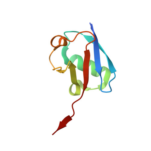
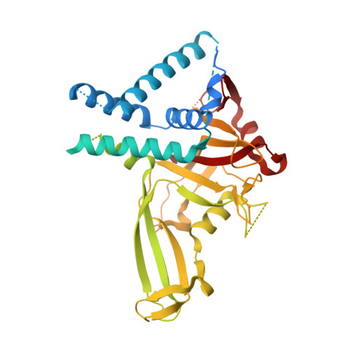
Cryo-EM reveals a mechanism of USP1 inhibition through a cryptic binding site.

Rennie, M.L.Arkinson, C.Chaugule, V.K.Walden, H.

(2022) Sci Adv 8: eabq6353-eabq6353

  • DOI: https://doi.org/10.1126/sciadv.abq6353
  • Primary Citation of Related Structures:  
    7ZH3, 7ZH4, 8A9J, 8A9K

  • PubMed Abstract: 

    Repair of DNA damage is critical to genomic integrity and frequently disrupted in cancers. Ubiquitin-specific protease 1 (USP1), a nucleus-localized deubiquitinase, lies at the interface of multiple DNA repair pathways and is a promising drug target for certain cancers. Although multiple inhibitors of this enzyme, including one in phase 1 clinical trials, have been established, their binding mode is unknown. Here, we use cryo-electron microscopy to study an assembled enzyme-substrate-inhibitor complex of USP1 and the well-established inhibitor, ML323. Achieving 2.5-Å resolution, with and without ML323, we find an unusual binding mode in which the inhibitor disrupts part of the hydrophobic core of USP1. The consequent conformational changes in the secondary structure lead to subtle rearrangements in the active site that underlie the mechanism of inhibition. These structures provide a platform for structure-based drug design targeting USP1.


  • Organizational Affiliation

    Institute of Molecular Cell and Systems Biology, College of Medical Veterinary and Life Sciences, University of Glasgow, Glasgow, UK.


Macromolecules
Find similar proteins by:  (by identity cutoff)  |  3D Structure
Entity ID: 1
MoleculeChains Sequence LengthOrganismDetailsImage
Ubiquitin-60S ribosomal protein L40A [auth C]80Homo sapiensMutation(s): 0 
Gene Names: UBA52UBCEP2
UniProt & NIH Common Fund Data Resources
Find proteins for P62987 (Homo sapiens)
Explore P62987 
Go to UniProtKB:  P62987
PHAROS:  P62987
GTEx:  ENSG00000221983 
Entity Groups  
Sequence Clusters30% Identity50% Identity70% Identity90% Identity95% Identity100% Identity
UniProt GroupP62987
Sequence Annotations
Expand
  • Reference Sequence
Find similar proteins by:  (by identity cutoff)  |  3D Structure
Entity ID: 2
MoleculeChains Sequence LengthOrganismDetailsImage
Ubiquitin carboxyl-terminal hydrolase 1B [auth D]786Homo sapiensMutation(s): 3 
Gene Names: USP1
EC: 3.4.19.12
UniProt & NIH Common Fund Data Resources
Find proteins for O94782 (Homo sapiens)
Explore O94782 
Go to UniProtKB:  O94782
PHAROS:  O94782
GTEx:  ENSG00000162607 
Entity Groups  
Sequence Clusters30% Identity50% Identity70% Identity90% Identity95% Identity100% Identity
UniProt GroupO94782
Sequence Annotations
Expand
  • Reference Sequence
Small Molecules
Ligands 2 Unique
IDChains Name / Formula / InChI Key2D Diagram3D Interactions
JDA (Subject of Investigation/LOI)
Query on JDA

Download Ideal Coordinates CCD File 
D
5-methyl-2-(2-propan-2-ylphenyl)-~{N}-[[4-(1,2,3-triazol-1-yl)phenyl]methyl]pyrimidin-4-amine
C23 H24 N6
VUIRVWPJNKZOSS-UHFFFAOYSA-N
ZN
Query on ZN

Download Ideal Coordinates CCD File 
C [auth D]ZINC ION
Zn
PTFCDOFLOPIGGS-UHFFFAOYSA-N
Experimental Data & Validation

Experimental Data

  • Method: ELECTRON MICROSCOPY
  • Resolution: 2.49 Å
  • Aggregation State: PARTICLE 
  • Reconstruction Method: SINGLE PARTICLE 
EM Software:
TaskSoftware PackageVersion
RECONSTRUCTIONcryoSPARC
MODEL REFINEMENTPHENIX1.19

Structure Validation

View Full Validation Report


Entry History & Funding Information

Deposition Data


Funding OrganizationLocationGrant Number
European Research Council (ERC)European UnionERC-2015-CoG-681582
Medical Research Council (MRC, United Kingdom)United KingdomMC_UU_12016/12

Revision History  (Full details and data files)

  • Version 1.0: 2022-10-12
    Type: Initial release