7ZBG

Human Topoisomerase II Beta ATPase ADP


Experimental Data Snapshot

  • Method: X-RAY DIFFRACTION
  • Resolution: 2.30 Å
  • R-Value Free: 0.259 
  • R-Value Work: 0.197 

wwPDB Validation   3D Report Full Report


Ligand Structure Quality Assessment 


This is version 1.2 of the entry. See complete history


Literature

A comprehensive structural analysis of the ATPase domain of human DNA topoisomerase II beta bound to AMPPNP, ADP, and the bisdioxopiperazine, ICRF193.

Ling, E.M.Basle, A.Cowell, I.G.van den Berg, B.Blower, T.R.Austin, C.A.

(2022) Structure 30: 1129-1145.e3

  • DOI: https://doi.org/10.1016/j.str.2022.05.009
  • Primary Citation of Related Structures:  
    7QFN, 7QFO, 7ZBG

  • PubMed Abstract: 

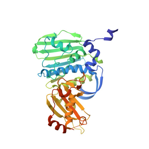
    Human topoisomerase II beta (TOP2B) modulates DNA topology using energy from ATP hydrolysis. To investigate the conformational changes that occur during ATP hydrolysis, we determined the X-ray crystallographic structures of the human TOP2B ATPase domain bound to AMPPNP or ADP at 1.9 Å and 2.6 Å resolution, respectively. The GHKL domains of both structures are similar, whereas the QTK loop within the transducer domain can move for product release. As TOP2B is the clinical target of bisdioxopiperazines, we also determined the structure of a TOP2B:ADP:ICRF193 complex to 2.3 Å resolution and identified key drug-binding residues. Biochemical characterization revealed the N-terminal strap reduces the rate of ATP hydrolysis. Mutagenesis demonstrated residue E103 as essential for ATP hydrolysis in TOP2B. Our data provide fundamental insights into the tertiary structure of the human TOP2B ATPase domain and a potential regulatory mechanism for ATP hydrolysis.


  • Organizational Affiliation

    Biosciences Institute, Newcastle University, Newcastle upon Tyne NE2 4HH, UK.


Macromolecules
Find similar proteins by:  (by identity cutoff)  |  3D Structure
Entity ID: 1
MoleculeChains Sequence LengthOrganismDetailsImage
DNA topoisomerase 2-beta400Homo sapiensMutation(s): 0 
Gene Names: TOP2B
EC: 5.6.2.2
UniProt & NIH Common Fund Data Resources
Find proteins for Q02880 (Homo sapiens)
Explore Q02880 
Go to UniProtKB:  Q02880
PHAROS:  Q02880
GTEx:  ENSG00000077097 
Entity Groups  
Sequence Clusters30% Identity50% Identity70% Identity90% Identity95% Identity100% Identity
UniProt GroupQ02880
Sequence Annotations
Expand
  • Reference Sequence
Small Molecules
Ligands 4 Unique
IDChains Name / Formula / InChI Key2D Diagram3D Interactions
ADP (Subject of Investigation/LOI)
Query on ADP

Download Ideal Coordinates CCD File 
B [auth A]ADENOSINE-5'-DIPHOSPHATE
C10 H15 N5 O10 P2
XTWYTFMLZFPYCI-KQYNXXCUSA-N
ITH (Subject of Investigation/LOI)
Query on ITH

Download Ideal Coordinates CCD File 
H [auth A]4-[(2S,3R)-3-[3,5-bis(oxidanylidene)piperazin-1-ium-1-yl]butan-2-yl]piperazin-4-ium-2,6-dione
C12 H20 N4 O4
OBYGAPWKTPDTAS-OCAPTIKFSA-P
SO4
Query on SO4

Download Ideal Coordinates CCD File 
C [auth A],
D [auth A],
E [auth A],
F [auth A]
SULFATE ION
O4 S
QAOWNCQODCNURD-UHFFFAOYSA-L
MG (Subject of Investigation/LOI)
Query on MG

Download Ideal Coordinates CCD File 
G [auth A]MAGNESIUM ION
Mg
JLVVSXFLKOJNIY-UHFFFAOYSA-N
Experimental Data & Validation

Experimental Data

  • Method: X-RAY DIFFRACTION
  • Resolution: 2.30 Å
  • R-Value Free: 0.259 
  • R-Value Work: 0.197 
  • Space Group: P 3 2 1
Unit Cell:
Length ( Å )Angle ( ˚ )
a = 103.4α = 90
b = 103.4β = 90
c = 67.02γ = 120
Software Package:
Software NamePurpose
REFMACrefinement
xia2data reduction
XDSdata reduction
Cootmodel building
MolProbitymodel building
pointlessdata scaling
Aimlessdata scaling
MOLREPphasing

Structure Validation

View Full Validation Report


Ligand Structure Quality Assessment 


Entry History & Funding Information

Deposition Data


Funding OrganizationLocationGrant Number
Not funded--

Revision History  (Full details and data files)

  • Version 1.0: 2022-05-25
    Type: Initial release
  • Version 1.1: 2022-11-30
    Changes: Database references
  • Version 1.2: 2024-01-31
    Changes: Data collection, Refinement description