7Z03

Endonuclease state of the E. coli Mre11-Rad50 (SbcCD) head complex bound to ADP and extended dsDNA


Experimental Data Snapshot

  • Method: ELECTRON MICROSCOPY
  • Resolution: 3.70 Å
  • Aggregation State: PARTICLE 
  • Reconstruction Method: SINGLE PARTICLE 

wwPDB Validation   3D Report Full Report


This is version 1.3 of the entry. See complete history


Literature

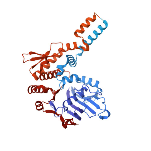
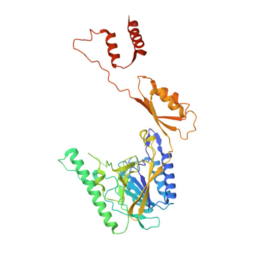
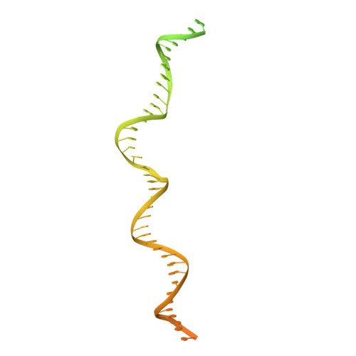
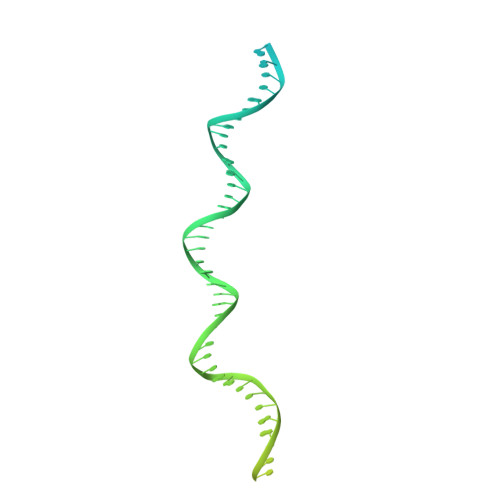
Structural mechanism of endonucleolytic processing of blocked DNA ends and hairpins by Mre11-Rad50.

Gut, F.Kashammer, L.Lammens, K.Bartho, J.D.Boggusch, A.M.van de Logt, E.Kessler, B.Hopfner, K.P.

(2022) Mol Cell 82: 3513-3522.e6

  • DOI: https://doi.org/10.1016/j.molcel.2022.07.019
  • Primary Citation of Related Structures:  
    7YZO, 7YZP, 7Z03

  • PubMed Abstract: 

    DNA double-strand breaks (DSBs) threaten genome stability and are linked to tumorigenesis in humans. Repair of DSBs requires the removal of attached proteins and hairpins through a poorly understood but physiologically critical endonuclease activity by the Mre11-Rad50 complex. Here, we report cryoelectron microscopy (cryo-EM) structures of the bacterial Mre11-Rad50 homolog SbcCD bound to a protein-blocked DNA end and a DNA hairpin. The structures reveal that Mre11-Rad50 bends internal DNA for endonucleolytic cleavage and show how internal DNA, DNA ends, and hairpins are processed through a similar ATP-regulated conformational state. Furthermore, Mre11-Rad50 is loaded onto blocked DNA ends with Mre11 pointing away from the block, explaining the distinct biochemistries of 3' → 5' exonucleolytic and endonucleolytic incision through the way Mre11-Rad50 interacts with diverse DNA ends. In summary, our results unify Mre11-Rad50's enigmatic nuclease diversity within a single structural framework and reveal how blocked DNA ends and hairpins are processed.


  • Organizational Affiliation

    Gene Center, Ludwig-Maximilians-Universität, 81377 Munich, Germany; Department of Biochemistry, Ludwig-Maximilians-Universität, 81377 Munich, Germany.


Macromolecules

Find similar proteins by:  (by identity cutoff)  |  3D Structure
Entity ID: 1
MoleculeChains Sequence LengthOrganismDetailsImage
Nuclease SbcCD subunit CA [auth C],
B [auth D]
1,048Escherichia coliMutation(s): 0 
Gene Names: sbcCrmuAb0397JW0387
UniProt
Find proteins for P13458 (Escherichia coli (strain K12))
Explore P13458 
Go to UniProtKB:  P13458
Entity Groups  
Sequence Clusters30% Identity50% Identity70% Identity90% Identity95% Identity100% Identity
UniProt GroupP13458
Sequence Annotations
Expand
  • Reference Sequence
Find similar proteins by:  (by identity cutoff)  |  3D Structure
Entity ID: 2
MoleculeChains Sequence LengthOrganismDetailsImage
Nuclease SbcCD subunit DC [auth A],
D [auth B]
407Escherichia coliMutation(s): 1 
Gene Names: sbcDyajAb0398JW0388
UniProt
Find proteins for P0AG76 (Escherichia coli (strain K12))
Explore P0AG76 
Go to UniProtKB:  P0AG76
Entity Groups  
Sequence Clusters30% Identity50% Identity70% Identity90% Identity95% Identity100% Identity
UniProt GroupP0AG76
Sequence Annotations
Expand
  • Reference Sequence
Find similar nucleic acids by:  (by identity cutoff)  |  3D Structure
Entity ID: 3
MoleculeChains LengthOrganismImage
DNA (39-MER)120synthetic construct
Sequence Annotations
Expand
  • Reference Sequence
Find similar nucleic acids by:  (by identity cutoff)  |  3D Structure
Entity ID: 4
MoleculeChains LengthOrganismImage
DNA (39-MER)120synthetic construct
Sequence Annotations
Expand
  • Reference Sequence
Small Molecules
Ligands 3 Unique
IDChains Name / Formula / InChI Key2D Diagram3D Interactions
ADP (Subject of Investigation/LOI)
Query on ADP

Download Ideal Coordinates CCD File 
G [auth C],
I [auth D]
ADENOSINE-5'-DIPHOSPHATE
C10 H15 N5 O10 P2
XTWYTFMLZFPYCI-KQYNXXCUSA-N
MN
Query on MN

Download Ideal Coordinates CCD File 
K [auth A],
L [auth A],
M [auth B],
N [auth B]
MANGANESE (II) ION
Mn
WAEMQWOKJMHJLA-UHFFFAOYSA-N
MG
Query on MG

Download Ideal Coordinates CCD File 
H [auth C],
J [auth D]
MAGNESIUM ION
Mg
JLVVSXFLKOJNIY-UHFFFAOYSA-N
Experimental Data & Validation

Experimental Data

  • Method: ELECTRON MICROSCOPY
  • Resolution: 3.70 Å
  • Aggregation State: PARTICLE 
  • Reconstruction Method: SINGLE PARTICLE 

Structure Validation

View Full Validation Report


Entry History & Funding Information

Deposition Data


Funding OrganizationLocationGrant Number
European Research Council (ERC)European Union--
German Research Foundation (DFG)Germany--

Revision History  (Full details and data files)

  • Version 1.0: 2022-08-17
    Type: Initial release
  • Version 1.1: 2022-08-24
    Changes: Source and taxonomy
  • Version 1.2: 2022-08-31
    Changes: Database references
  • Version 1.3: 2022-09-28
    Changes: Database references