7YTE

crystal structure of human FcmR-D1 bound to IgM C4-domain


Experimental Data Snapshot

  • Method: X-RAY DIFFRACTION
  • Resolution: 3.00 Å
  • R-Value Free: 0.269 
  • R-Value Work: 0.230 
  • R-Value Observed: 0.232 

wwPDB Validation   3D Report Full Report


This is version 2.2 of the entry. See complete history


Literature

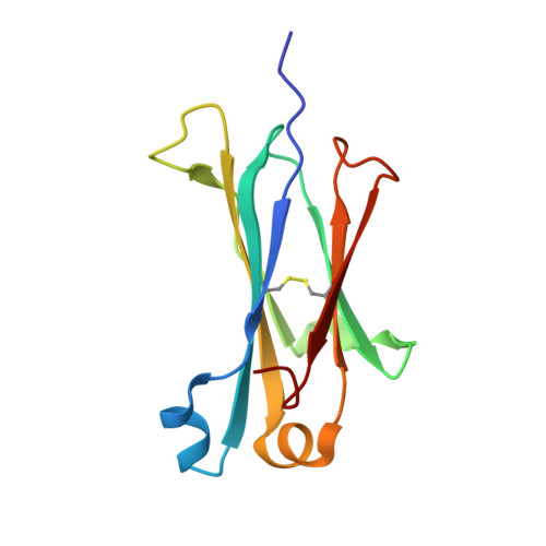
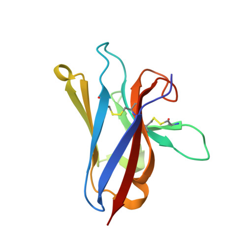
Immunoglobulin M perception by Fc mu R.

Li, Y.Shen, H.Zhang, R.Ji, C.Wang, Y.Su, C.Xiao, J.

(2023) Nature 615: 907-912

  • DOI: https://doi.org/10.1038/s41586-023-05835-w
  • Primary Citation of Related Structures:  
    7YSG, 7YTC, 7YTD, 7YTE

  • PubMed Abstract: 

    Immunoglobulin M (IgM) is the first antibody to emerge during embryonic development and the humoral immune response 1 . IgM can exist in several distinct forms, including monomeric, membrane-bound IgM within the B cell receptor (BCR) complex, pentameric and hexameric IgM in serum and secretory IgM on the mucosal surface. FcμR, the only IgM-specific receptor in mammals, recognizes different forms of IgM to regulate diverse immune responses 2-5 . However, the underlying molecular mechanisms remain unknown. Here we delineate the structural basis of the FcμR-IgM interaction by crystallography and cryo-electron microscopy. We show that two FcμR molecules interact with a Fcμ-Cμ4 dimer, suggesting that FcμR can bind to membrane-bound IgM with a 2:1 stoichiometry. Further analyses reveal that FcμR-binding sites are accessible in the context of IgM BCR. By contrast, pentameric IgM can recruit four FcμR molecules to bind on the same side and thereby facilitate the formation of an FcμR oligomer. One of these FcμR molecules occupies the binding site of the secretory component. Nevertheless, four FcμR molecules bind to the other side of secretory component-containing secretory IgM, consistent with the function of FcμR in the retrotransport of secretory IgM. These results reveal intricate mechanisms of IgM perception by FcμR.


  • Organizational Affiliation

    State Key Laboratory of Protein and Plant Gene Research, School of Life Sciences, Peking University, Beijing, P. R. China.


Macromolecules
Find similar proteins by:  (by identity cutoff)  |  3D Structure
Entity ID: 1
MoleculeChains Sequence LengthOrganismDetailsImage
Ig mu chain C region secreted form
A, B
112Homo sapiensMutation(s): 0 
Entity Groups  
Sequence Clusters30% Identity50% Identity70% Identity90% Identity95% Identity100% Identity
Sequence Annotations
Expand
  • Reference Sequence
Find similar proteins by:  (by identity cutoff)  |  3D Structure
Entity ID: 2
MoleculeChains Sequence LengthOrganismDetailsImage
Fas apoptotic inhibitory molecule 3C [auth D],
D [auth C]
107Homo sapiensMutation(s): 0 
Gene Names: FCMR
UniProt & NIH Common Fund Data Resources
Find proteins for O60667 (Homo sapiens)
Explore O60667 
Go to UniProtKB:  O60667
PHAROS:  O60667
GTEx:  ENSG00000162894 
Entity Groups  
Sequence Clusters30% Identity50% Identity70% Identity90% Identity95% Identity100% Identity
UniProt GroupO60667
Sequence Annotations
Expand
  • Reference Sequence
Experimental Data & Validation

Experimental Data

  • Method: X-RAY DIFFRACTION
  • Resolution: 3.00 Å
  • R-Value Free: 0.269 
  • R-Value Work: 0.230 
  • R-Value Observed: 0.232 
  • Space Group: P 3
Unit Cell:
Length ( Å )Angle ( ˚ )
a = 85.718α = 90
b = 85.718β = 90
c = 60.676γ = 120
Software Package:
Software NamePurpose
PHENIXrefinement
PDB_EXTRACTdata extraction
HKL-2000data reduction
HKL-2000data scaling
PHASERphasing

Structure Validation

View Full Validation Report


Entry History & Funding Information

Deposition Data


Funding OrganizationLocationGrant Number
National Science Foundation (NSF, China)China--

Revision History  (Full details and data files)

  • Version 1.0: 2023-02-01
    Type: Initial release
  • Version 2.0: 2023-02-08
    Changes: Advisory, Atomic model, Data collection, Database references, Derived calculations, Experimental preparation, Polymer sequence, Refinement description, Source and taxonomy, Structure summary
  • Version 2.1: 2023-08-16
    Changes: Data collection, Database references, Refinement description
  • Version 2.2: 2023-11-29
    Changes: Refinement description