7WAB

Crystal structure of the prolyl endoprotease, PEP, from Aspergillus niger


Experimental Data Snapshot

  • Method: X-RAY DIFFRACTION
  • Resolution: 1.75 Å
  • R-Value Free: 0.193 
  • R-Value Work: 0.164 
  • R-Value Observed: 0.165 

wwPDB Validation   3D Report Full Report


Ligand Structure Quality Assessment 


This is version 1.1 of the entry. See complete history


Literature

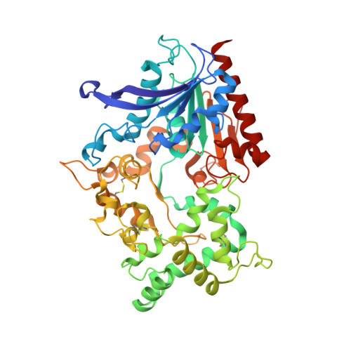
Crystal structure and substrate recognition mechanism of the prolyl endoprotease PEP from Aspergillus niger.

Miyazono, K.I.Kubota, K.Takahashi, K.Tanokura, M.

(2022) Biochem Biophys Res Commun 591: 76-81

  • DOI: https://doi.org/10.1016/j.bbrc.2021.12.114
  • Primary Citation of Related Structures:  
    7WAB

  • PubMed Abstract: 

    Proteases are enzymes that are not only essential for life but also industrially important. Understanding the substrate recognition mechanisms of proteases is important to enhance the use of proteases. The fungus Aspergillus produces a wide variety of proteases, including PEP, which is a prolyl endoprotease from A. niger. Although PEP exhibits amino acid sequence similarity to the serine peptidase family S28 proteins (PRCP and DPP7) that recognize Pro-X bonds in the terminal regions of peptides, PEP recognizes Pro-X bonds not only in peptides but also in proteins. To reveal the structural basis of the prolyl endoprotease activity of PEP, we determined the structure of PEP by X-ray crystallography at a resolution of 1.75 Å. The PEP structure shows that PEP has a wide-open catalytic pocket compared to its homologs. The characteristic catalytic pocket structure of PEP is predicted to be important for the recognition of protein substrates.


  • Organizational Affiliation

    Department of Applied Biological Chemistry, Graduate School of Agricultural and Life Sciences, The University of Tokyo, 1-1-1 Yayoi Bunkyo-ku, Tokyo, 113-8657, Japan.


Macromolecules
Find similar proteins by:  (by identity cutoff)  |  3D Structure
Entity ID: 1
MoleculeChains Sequence LengthOrganismDetailsImage
COMPASS (Complex proteins associated with Set1p) component shg1 family protein484Aspergillus nigerMutation(s): 0 
UniProt
Find proteins for A0A3F3RXI7 (Aspergillus niger)
Explore A0A3F3RXI7 
Go to UniProtKB:  A0A3F3RXI7
Entity Groups  
Sequence Clusters30% Identity50% Identity70% Identity90% Identity95% Identity100% Identity
UniProt GroupA0A3F3RXI7
Sequence Annotations
Expand
  • Reference Sequence
Oligosaccharides

Help

Entity ID: 2
MoleculeChains Length2D Diagram Glycosylation3D Interactions
alpha-D-mannopyranose-(1-2)-alpha-D-mannopyranose-(1-2)-alpha-D-mannopyranose-(1-3)-beta-D-mannopyranose-(1-4)-2-acetamido-2-deoxy-beta-D-glucopyranose-(1-4)-2-acetamido-2-deoxy-beta-D-glucopyranoseB [auth C]6N-Glycosylation
Glycosylation Resources
GlyTouCan:  G01760ZU
GlyCosmos:  G01760ZU
GlyGen:  G01760ZU
Entity ID: 3
MoleculeChains Length2D Diagram Glycosylation3D Interactions
2-acetamido-2-deoxy-beta-D-glucopyranose-(1-4)-2-acetamido-2-deoxy-beta-D-glucopyranoseC [auth D]2N-Glycosylation
Glycosylation Resources
GlyTouCan:  G42666HT
GlyCosmos:  G42666HT
GlyGen:  G42666HT
Small Molecules
Experimental Data & Validation

Experimental Data

  • Method: X-RAY DIFFRACTION
  • Resolution: 1.75 Å
  • R-Value Free: 0.193 
  • R-Value Work: 0.164 
  • R-Value Observed: 0.165 
  • Space Group: P 4 21 2
Unit Cell:
Length ( Å )Angle ( ˚ )
a = 144.517α = 90
b = 144.517β = 90
c = 56.542γ = 90
Software Package:
Software NamePurpose
Aimlessdata scaling
PHENIXrefinement
PDB_EXTRACTdata extraction
XDSdata reduction
MOLREPphasing

Structure Validation

View Full Validation Report


Ligand Structure Quality Assessment 


Entry History & Funding Information

Deposition Data


Funding OrganizationLocationGrant Number
Ministry of Education, Culture, Sports, Science and Technology (Japan)Japan--

Revision History  (Full details and data files)

  • Version 1.0: 2022-01-12
    Type: Initial release
  • Version 1.1: 2022-02-16
    Changes: Database references