


Crystal structure of siderophore binding protein VatD from Vibrio vulnificus M2799 complexed with Desferal
Tomoo, K., Miyamoto, K.To be published.
Experimental Data Snapshot
Starting Model: experimental
View more details
Entity ID: 1 | |||||
|---|---|---|---|---|---|
| Molecule | Chains | Sequence Length | Organism | Details | Image |
| Putative periplasm binding protein | 280 | Vibrio vulnificus | Mutation(s): 0 Gene Names: fhuD | 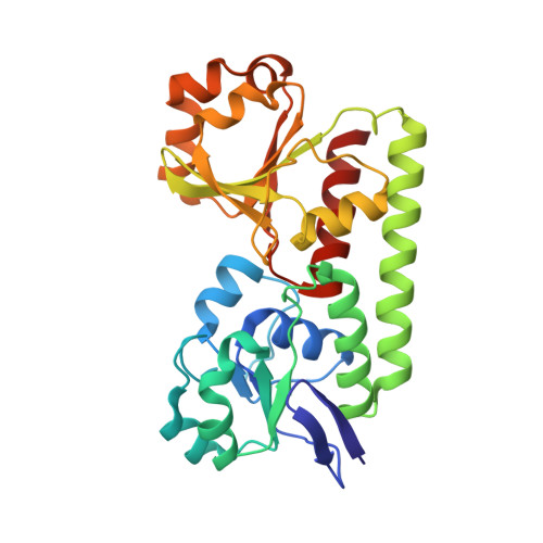 | |
UniProt | |||||
Entity Groups | |||||
| Sequence Clusters | 30% Identity50% Identity70% Identity90% Identity95% Identity100% Identity | ||||
| UniProt Group | Q845T3 | ||||
Sequence AnnotationsExpand | |||||
Reference Sequence | |||||
| Ligands 3 Unique | |||||
|---|---|---|---|---|---|
| ID | Chains | Name / Formula / InChI Key | 2D Diagram | 3D Interactions | |
| KTY (Subject of Investigation/LOI) Download:Ideal Coordinates CCD File | B [auth A] | desferrioxamine B C25 H48 N6 O8 UBQYURCVBFRUQT-UHFFFAOYSA-N |  | ||
| FE Download:Ideal Coordinates CCD File | C [auth A], D [auth A] | FE (III) ION Fe VTLYFUHAOXGGBS-UHFFFAOYSA-N |  | ||
| MG Download:Ideal Coordinates CCD File | E [auth A], F [auth A] | MAGNESIUM ION Mg JLVVSXFLKOJNIY-UHFFFAOYSA-N |  | ||
| Length ( Å ) | Angle ( ˚ ) |
|---|---|
| a = 34.8 | α = 90 |
| b = 57.89 | β = 95.02 |
| c = 62.7 | γ = 90 |
| Software Name | Purpose |
|---|---|
| REFMAC | refinement |
| CrystalClear | data reduction |
| CrystalClear | data scaling |
| MOLREP | phasing |
| Funding Organization | Location | Grant Number |
|---|---|---|
| Not funded | -- |


