7VSA

E. coli Ribonuclease HI in complex with two Mg2+


Experimental Data Snapshot

  • Method: X-RAY DIFFRACTION
  • Resolution: 1.76 Å
  • R-Value Free: 0.233 
  • R-Value Work: 0.201 
  • R-Value Observed: 0.202 

wwPDB Validation   3D Report Full Report


This is version 1.2 of the entry. See complete history


Literature

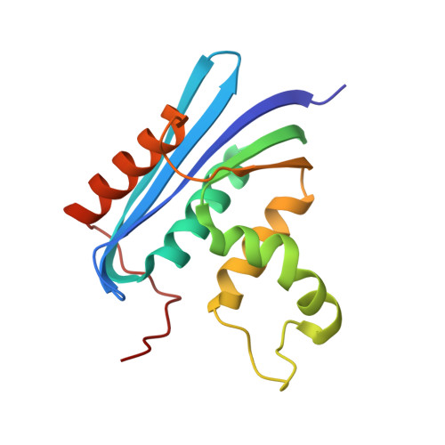
Pivotal role of a conserved histidine in Escherichia coli ribonuclease HI as proposed by X-ray crystallography.

Liao, Z.Oyama, T.Kitagawa, Y.Katayanagi, K.Morikawa, K.Oda, M.

(2022) Acta Crystallogr D Struct Biol 78: 390-398

  • DOI: https://doi.org/10.1107/S2059798322000870
  • Primary Citation of Related Structures:  
    7VSA, 7VSB, 7VSC, 7VSD, 7VSE

  • PubMed Abstract: 

    The ribonuclease (RNase) H family of enzymes catalyze the specific cleavage of RNA strands of RNA/DNA hybrid duplexes and play an important role in DNA replication and repair. Since the first report of the crystal structure of RNase HI, its catalytic mechanisms, which require metal ions, have been discussed based on numerous structural and functional analyses, including X-ray crystallography. In contrast, the function of the conserved histidine residue (His124 in Escherichia coli) in the flexible loop around the active site remains poorly understood, although an important role was suggested by NMR analyses. Here, novel high-resolution X-ray crystal structures of E. coli RNase HI are described, with a particular focus on the interactions of divalent cations with His124 oriented towards the active site. The enzyme-Mg 2+ complex contains two metal ions in the active site, one of which has previously been observed. The second ion lies alongside the first and binds to His124 in an octahedral coordination scheme. In the enzyme-Zn 2+ complex a single metal ion was found to bind to the active site, showing a tetrahedral coordination geometry with the surrounding atoms, including His124. These results provide structural evidence that His124 plays a crucial role in the catalytic activity of RNase HI by interacting weakly and transiently with metal ions in the catalytic center.


  • Organizational Affiliation

    Graduate School of Life and Environmental Sciences, Kyoto Prefectural University, 1-5 Hangi-cho, Shimogamo, Sakyo-ku, Kyoto 606-8522, Japan.


Macromolecules
Find similar proteins by:  (by identity cutoff)  |  3D Structure
Entity ID: 1
MoleculeChains Sequence LengthOrganismDetailsImage
Ribonuclease HI
A, B
155Escherichia coli K-12Mutation(s): 0 
Gene Names: rnhAdasFherArnhsdrAb0214JW0204
EC: 3.1.26.4
UniProt
Find proteins for P0A7Y4 (Escherichia coli (strain K12))
Explore P0A7Y4 
Go to UniProtKB:  P0A7Y4
Entity Groups  
Sequence Clusters30% Identity50% Identity70% Identity90% Identity95% Identity100% Identity
UniProt GroupP0A7Y4
Sequence Annotations
Expand
  • Reference Sequence
Small Molecules
Ligands 3 Unique
IDChains Name / Formula / InChI Key2D Diagram3D Interactions
SO4
Query on SO4

Download Ideal Coordinates CCD File 
G [auth A],
H [auth A],
K [auth B]
SULFATE ION
O4 S
QAOWNCQODCNURD-UHFFFAOYSA-L
GOL
Query on GOL

Download Ideal Coordinates CCD File 
C [auth A],
D [auth A]
GLYCEROL
C3 H8 O3
PEDCQBHIVMGVHV-UHFFFAOYSA-N
MG (Subject of Investigation/LOI)
Query on MG

Download Ideal Coordinates CCD File 
E [auth A],
F [auth A],
I [auth B],
J [auth B]
MAGNESIUM ION
Mg
JLVVSXFLKOJNIY-UHFFFAOYSA-N
Experimental Data & Validation

Experimental Data

  • Method: X-RAY DIFFRACTION
  • Resolution: 1.76 Å
  • R-Value Free: 0.233 
  • R-Value Work: 0.201 
  • R-Value Observed: 0.202 
  • Space Group: P 2 2 21
Unit Cell:
Length ( Å )Angle ( ˚ )
a = 61.639α = 90
b = 64.288β = 90
c = 80.288γ = 90
Software Package:
Software NamePurpose
PHENIXrefinement
XDSdata reduction
Aimlessdata scaling
PHASERphasing

Structure Validation

View Full Validation Report


Entry History & Funding Information

Deposition Data


Funding OrganizationLocationGrant Number
Japan Society for the Promotion of Science (JSPS)Japan21H02120
Japan Science and TechnologyJapanJPMJTM20NM

Revision History  (Full details and data files)

  • Version 1.0: 2022-03-02
    Type: Initial release
  • Version 1.1: 2022-03-16
    Changes: Database references
  • Version 1.2: 2023-11-29
    Changes: Data collection, Refinement description