7UJ0

ClpAP complex bound to ClpS N-terminal extension, class IIIb


Experimental Data Snapshot

  • Method: ELECTRON MICROSCOPY
  • Resolution: 3.26 Å
  • Aggregation State: PARTICLE 
  • Reconstruction Method: SINGLE PARTICLE 

wwPDB Validation   3D Report Full Report


This is version 1.2 of the entry. See complete history


Literature

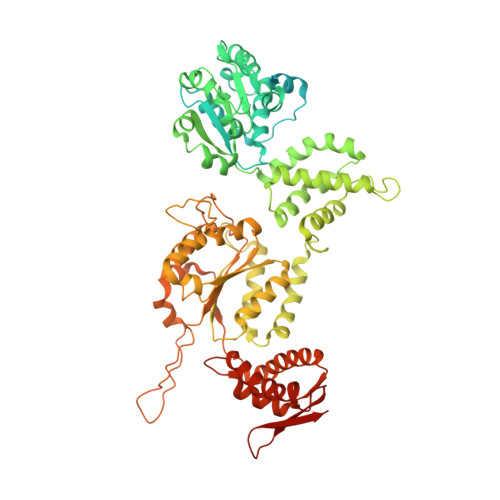
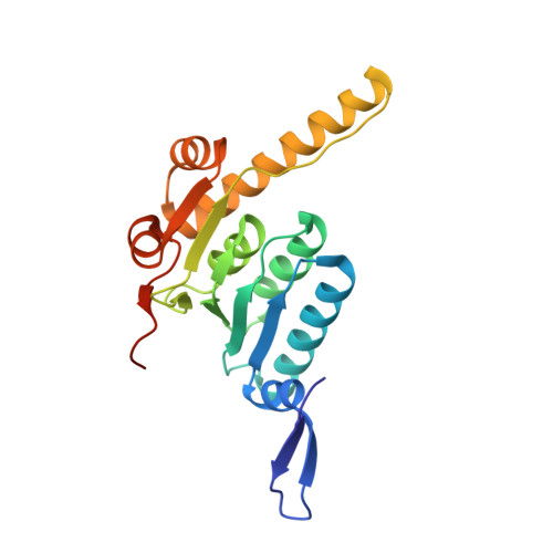
AAA+ protease-adaptor structures reveal altered conformations and ring specialization.

Kim, S.Fei, X.Sauer, R.T.Baker, T.A.

(2022) Nat Struct Mol Biol 29: 1068-1079

  • DOI: https://doi.org/10.1038/s41594-022-00850-3
  • Primary Citation of Related Structures:  
    7UIV, 7UIW, 7UIX, 7UIY, 7UIZ, 7UJ0

  • PubMed Abstract: 

    ClpAP, a two-ring AAA+ protease, degrades N-end-rule proteins bound by the ClpS adaptor. Here we present high-resolution cryo-EM structures of Escherichia coli ClpAPS complexes, showing how ClpA pore loops interact with the ClpS N-terminal extension (NTE), which is normally intrinsically disordered. In two classes, the NTE is bound by a spiral of pore-1 and pore-2 loops in a manner similar to substrate-polypeptide binding by many AAA+ unfoldases. Kinetic studies reveal that pore-2 loops of the ClpA D1 ring catalyze the protein remodeling required for substrate delivery by ClpS. In a third class, D2 pore-1 loops are rotated, tucked away from the channel and do not bind the NTE, demonstrating asymmetry in engagement by the D1 and D2 rings. These studies show additional structures and functions for key AAA+ elements. Pore-loop tucking may be used broadly by AAA+ unfoldases, for example, during enzyme pausing/unloading.


  • Organizational Affiliation

    Department of Biology, Massachusetts Institute of Technology, Cambridge, MA, USA.


Macromolecules
Find similar proteins by:  (by identity cutoff)  |  3D Structure
Entity ID: 1
MoleculeChains Sequence LengthOrganismDetailsImage
ATP-dependent Clp protease ATP-binding subunit ClpA
A, B, C, D, E
A, B, C, D, E, F
758Escherichia coliMutation(s): 0 
Gene Names: clpAAB05_1038
UniProt
Find proteins for P0ABH9 (Escherichia coli (strain K12))
Explore P0ABH9 
Go to UniProtKB:  P0ABH9
Entity Groups  
Sequence Clusters30% Identity50% Identity70% Identity90% Identity95% Identity100% Identity
UniProt GroupP0ABH9
Sequence Annotations
Expand
  • Reference Sequence
Find similar proteins by:  (by identity cutoff)  |  3D Structure
Entity ID: 2
MoleculeChains Sequence LengthOrganismDetailsImage
ATP-dependent Clp protease adapter protein ClpSG [auth S]106Escherichia coliMutation(s): 0 
Gene Names: clpSECVG_02246
UniProt
Find proteins for P0A8Q6 (Escherichia coli (strain K12))
Explore P0A8Q6 
Go to UniProtKB:  P0A8Q6
Entity Groups  
Sequence Clusters30% Identity50% Identity70% Identity90% Identity95% Identity100% Identity
UniProt GroupP0A8Q6
Sequence Annotations
Expand
  • Reference Sequence
Find similar proteins by:  (by identity cutoff)  |  3D Structure
Entity ID: 3
MoleculeChains Sequence LengthOrganismDetailsImage
ATP-dependent Clp protease proteolytic subunit
H, I, J, K, L
H, I, J, K, L, M, N
201Escherichia coliMutation(s): 0 
Gene Names: clpP
EC: 3.4.21.92
UniProt
Find proteins for P0A6G7 (Escherichia coli (strain K12))
Explore P0A6G7 
Go to UniProtKB:  P0A6G7
Entity Groups  
Sequence Clusters30% Identity50% Identity70% Identity90% Identity95% Identity100% Identity
UniProt GroupP0A6G7
Sequence Annotations
Expand
  • Reference Sequence
Small Molecules
Ligands 3 Unique
IDChains Name / Formula / InChI Key2D Diagram3D Interactions
AGS (Subject of Investigation/LOI)
Query on AGS

Download Ideal Coordinates CCD File 
AA [auth D]
BA [auth D]
O [auth A]
S [auth B]
T [auth B]
AA [auth D],
BA [auth D],
O [auth A],
S [auth B],
T [auth B],
W [auth C],
X [auth C]
PHOSPHOTHIOPHOSPHORIC ACID-ADENYLATE ESTER
C10 H16 N5 O12 P3 S
NLTUCYMLOPLUHL-KQYNXXCUSA-N
ADP (Subject of Investigation/LOI)
Query on ADP

Download Ideal Coordinates CCD File 
EA [auth E],
FA [auth E],
HA [auth F],
IA [auth F],
P [auth A]
ADENOSINE-5'-DIPHOSPHATE
C10 H15 N5 O10 P2
XTWYTFMLZFPYCI-KQYNXXCUSA-N
MG (Subject of Investigation/LOI)
Query on MG

Download Ideal Coordinates CCD File 
CA [auth D]
DA [auth D]
GA [auth E]
Q [auth A]
R [auth A]
CA [auth D],
DA [auth D],
GA [auth E],
Q [auth A],
R [auth A],
U [auth B],
V [auth B],
Y [auth C],
Z [auth C]
MAGNESIUM ION
Mg
JLVVSXFLKOJNIY-UHFFFAOYSA-N
Experimental Data & Validation

Experimental Data

  • Method: ELECTRON MICROSCOPY
  • Resolution: 3.26 Å
  • Aggregation State: PARTICLE 
  • Reconstruction Method: SINGLE PARTICLE 
EM Software:
TaskSoftware PackageVersion
MODEL REFINEMENTPHENIX
MODEL REFINEMENTCoot
RECONSTRUCTIONPHENIX

Structure Validation

View Full Validation Report


Entry History & Funding Information

Deposition Data


Funding OrganizationLocationGrant Number
National Institutes of Health/National Institute of General Medical Sciences (NIH/NIGMS)United States--
Howard Hughes Medical Institute (HHMI)United States--

Revision History  (Full details and data files)

  • Version 1.0: 2022-11-09
    Type: Initial release
  • Version 1.1: 2022-11-16
    Changes: Database references
  • Version 1.2: 2022-11-30
    Changes: Database references