7U8K

Magic Angle Spinning NMR Structure of Human Cofilin-2 Assembled on Actin Filaments


Experimental Data Snapshot

  • Method: SOLID-STATE NMR
  • Conformers Calculated: 
  • Conformers Submitted: 
  • Selection Criteria: all calculated structures submitted 

wwPDB Validation   3D Report Full Report

Currently 7U8K does not have a validation slider image.


This is version 1.1 of the entry. See complete history


Literature

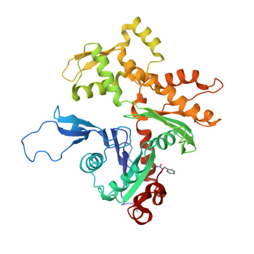
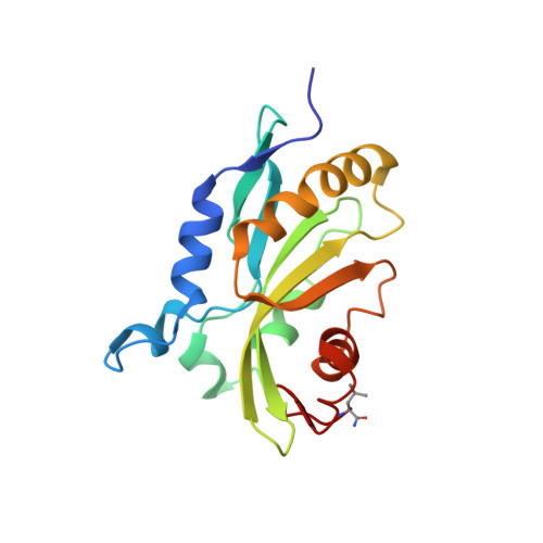
Magic angle spinning NMR structure of human cofilin-2 assembled on actin filaments reveals isoform-specific conformation and binding mode.

Kraus, J.Russell, R.W.Kudryashova, E.Xu, C.Katyal, N.Perilla, J.R.Kudryashov, D.S.Polenova, T.

(2022) Nat Commun 13: 2114-2114

  • DOI: https://doi.org/10.1038/s41467-022-29595-9
  • Primary Citation of Related Structures:  
    7M0G, 7U8K

  • PubMed Abstract: 

    Actin polymerization dynamics regulated by actin-binding proteins are essential for various cellular functions. The cofilin family of proteins are potent regulators of actin severing and filament disassembly. The structural basis for cofilin-isoform-specific severing activity is poorly understood as their high-resolution structures in complex with filamentous actin (F-actin) are lacking. Here, we present the atomic-resolution structure of the muscle-tissue-specific isoform, cofilin-2 (CFL2), assembled on ADP-F-actin, determined by magic-angle-spinning (MAS) NMR spectroscopy and data-guided molecular dynamics (MD) simulations. We observe an isoform-specific conformation for CFL2. This conformation is the result of a unique network of hydrogen bonding interactions within the α2 helix containing the non-conserved residue, Q26. Our results indicate F-site interactions that are specific between CFL2 and ADP-F-actin, revealing mechanistic insights into isoform-dependent F-actin disassembly.


  • Organizational Affiliation

    Department of Chemistry and Biochemistry, University of Delaware, Newark, DE, 19716, United States.


Macromolecules
Find similar proteins by:  (by identity cutoff)  |  3D Structure
Entity ID: 1
MoleculeChains Sequence LengthOrganismDetailsImage
Actin, alpha skeletal muscle
A, B, C, D, E
A, B, C, D, E, F, G, H, I, J
377Oryctolagus cuniculusMutation(s): 2 
Gene Names: ACTA1ACTA
EC: 3.6.4
UniProt
Find proteins for P68135 (Oryctolagus cuniculus)
Explore P68135 
Go to UniProtKB:  P68135
Entity Groups  
Sequence Clusters30% Identity50% Identity70% Identity90% Identity95% Identity100% Identity
UniProt GroupP68135
Sequence Annotations
Expand
  • Reference Sequence
Find similar proteins by:  (by identity cutoff)  |  3D Structure
Entity ID: 2
MoleculeChains Sequence LengthOrganismDetailsImage
Cofilin-2
K, L, M, N, O
K, L, M, N, O, P, Q, R
168Homo sapiensMutation(s): 2 
Gene Names: CFL2
UniProt & NIH Common Fund Data Resources
Find proteins for Q9Y281 (Homo sapiens)
Explore Q9Y281 
Go to UniProtKB:  Q9Y281
PHAROS:  Q9Y281
GTEx:  ENSG00000165410 
Entity Groups  
Sequence Clusters30% Identity50% Identity70% Identity90% Identity95% Identity100% Identity
UniProt GroupQ9Y281
Sequence Annotations
Expand
  • Reference Sequence
Experimental Data & Validation

Experimental Data

  • Method: SOLID-STATE NMR
  • Conformers Calculated: 
  • Conformers Submitted: 
  • Selection Criteria: all calculated structures submitted 

Structure Validation

View Full Validation Report

Currently 7U8K does not have a validation slider image.


Entry History & Funding Information

Deposition Data


Funding OrganizationLocationGrant Number
National Science Foundation (NSF, United States)United States1247394
National Science Foundation (NSF, United States)United StatesCHE0959496
National Institutes of Health/National Institute of General Medical Sciences (NIH/NIGMS)United StatesP30GM110758

Revision History  (Full details and data files)

  • Version 1.0: 2023-08-16
    Type: Initial release
  • Version 1.1: 2024-02-28
    Changes: Database references