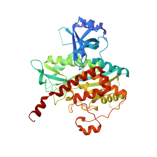
7TDP

Structure of Paenibacillus polymyxa GS bound to Met-Sox-P-ADP (Transition state complex) to 1.98 Angstom

  • Classification: LIGASE
  • Organism(s): Paenibacillus polymyxa
  • Expression System: Escherichia coli
  • Mutation(s): No 

  • Deposited: 2022-01-02 Released: 2022-06-29 
  • Deposition Author(s): Schumacher, M.A.
  • Funding Organization(s): National Institutes of Health/National Institute of General Medical Sciences (NIH/NIGMS)

Experimental Data Snapshot

  • Method: X-RAY DIFFRACTION
  • Resolution: 1.98 Å
  • R-Value Free: 0.201 
  • R-Value Work: 0.171 
  • R-Value Observed: 0.171 

Starting Model: experimental
View more details

wwPDB Validation   3D Report Full Report


Ligand Structure Quality Assessment 


This is version 1.2 of the entry. See complete history


Literature

Molecular dissection of the glutamine synthetase-GlnR nitrogen regulatory circuitry in Gram-positive bacteria.

Travis, B.A.Peck, J.V.Salinas, R.Dopkins, B.Lent, N.Nguyen, V.D.Borgnia, M.J.Brennan, R.G.Schumacher, M.A.

(2022) Nat Commun 13: 3793-3793

  • DOI: https://doi.org/10.1038/s41467-022-31573-0
  • Primary Citation of Related Structures:  
    7TDP, 7TDV, 7TEA, 7TEC, 7TEN, 7TF6, 7TF7, 7TF9, 7TFA, 7TFB, 7TFC, 7TFD, 7TFE

  • PubMed Abstract: 

    How bacteria sense and respond to nitrogen levels are central questions in microbial physiology. In Gram-positive bacteria, nitrogen homeostasis is controlled by an operon encoding glutamine synthetase (GS), a dodecameric machine that assimilates ammonium into glutamine, and the GlnR repressor. GlnR detects nitrogen excess indirectly by binding glutamine-feedback-inhibited-GS (FBI-GS), which activates its transcription-repression function. The molecular mechanisms behind this regulatory circuitry, however, are unknown. Here we describe biochemical and structural analyses of GS and FBI-GS-GlnR complexes from pathogenic and non-pathogenic Gram-positive bacteria. The structures show FBI-GS binds the GlnR C-terminal domain within its active-site cavity, juxtaposing two GlnR monomers to form a DNA-binding-competent GlnR dimer. The FBI-GS-GlnR interaction stabilizes the inactive GS conformation. Strikingly, this interaction also favors a remarkable dodecamer to tetradecamer transition in some GS, breaking the paradigm that all bacterial GS are dodecamers. These data thus unveil unique structural mechanisms of transcription and enzymatic regulation.


  • Organizational Affiliation

    Department of Biochemistry, 307 Research Dr., Box 3711, Duke University Medical Center, Durham, NC, 27710, USA.


Macromolecules
Find similar proteins by:  (by identity cutoff)  |  3D Structure
Entity ID: 1
MoleculeChains Sequence LengthOrganismDetailsImage
Glutamine synthetaseA,
B [auth C],
C [auth B]
442Paenibacillus polymyxaMutation(s): 0 
Gene Names: glnA2LK13_01575NCTC10343_02989
EC: 6.3.1.2
UniProt
Find proteins for A0A0F0G8G2 (Paenibacillus polymyxa)
Explore A0A0F0G8G2 
Go to UniProtKB:  A0A0F0G8G2
Entity Groups  
Sequence Clusters30% Identity50% Identity70% Identity90% Identity95% Identity100% Identity
UniProt GroupA0A0F0G8G2
Sequence Annotations
Expand
  • Reference Sequence
Small Molecules
Ligands 3 Unique
IDChains Name / Formula / InChI Key2D Diagram3D Interactions
ADP
Query on ADP

Download Ideal Coordinates CCD File 
D [auth A],
I [auth C],
N [auth B]
ADENOSINE-5'-DIPHOSPHATE
C10 H15 N5 O10 P2
XTWYTFMLZFPYCI-KQYNXXCUSA-N
P3S (Subject of Investigation/LOI)
Query on P3S

Download Ideal Coordinates CCD File 
E [auth A],
J [auth C],
O [auth B]
L-METHIONINE-S-SULFOXIMINE PHOSPHATE
C5 H13 N2 O6 P S
QQFOFBSCSWFFPB-NMAPHRJESA-N
MG
Query on MG

Download Ideal Coordinates CCD File 
F [auth A]
G [auth A]
H [auth A]
K [auth C]
L [auth C]
F [auth A],
G [auth A],
H [auth A],
K [auth C],
L [auth C],
M [auth C],
P [auth B],
Q [auth B],
R [auth B]
MAGNESIUM ION
Mg
JLVVSXFLKOJNIY-UHFFFAOYSA-N
Experimental Data & Validation

Experimental Data

  • Method: X-RAY DIFFRACTION
  • Resolution: 1.98 Å
  • R-Value Free: 0.201 
  • R-Value Work: 0.171 
  • R-Value Observed: 0.171 
  • Space Group: P 42 21 2
Unit Cell:
Length ( Å )Angle ( ˚ )
a = 164.009α = 90
b = 164.009β = 90
c = 141.528γ = 90
Software Package:
Software NamePurpose
XDSdata reduction
SCALAdata scaling
PHASERphasing
PHENIXrefinement
PDB_EXTRACTdata extraction

Structure Validation

View Full Validation Report


Ligand Structure Quality Assessment 


Entry History & Funding Information

Deposition Data


Funding OrganizationLocationGrant Number
National Institutes of Health/National Institute of General Medical Sciences (NIH/NIGMS)United StatesR35GM130290

Revision History  (Full details and data files)

  • Version 1.0: 2022-06-29
    Type: Initial release
  • Version 1.1: 2022-07-13
    Changes: Database references
  • Version 1.2: 2023-10-18
    Changes: Data collection, Refinement description