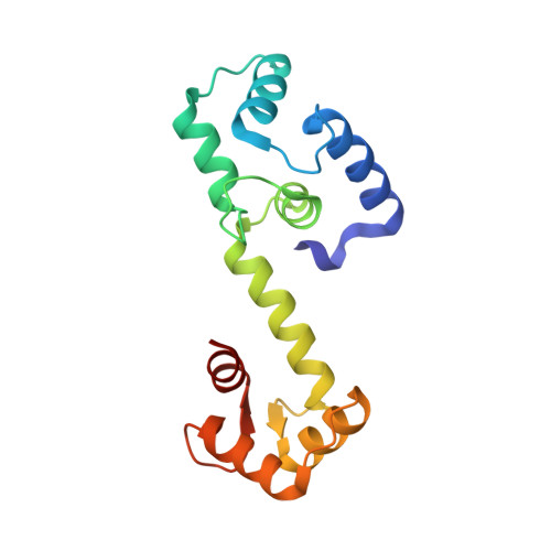
Site-selective photocatalytic functionalization of peptides and proteins at selenocysteine.
Dowman, L.J., Kulkarni, S.S., Alegre-Requena, J.V., Giltrap, A.M., Norman, A.R., Sharma, A., Gallegos, L.C., Mackay, A.S., Welegedara, A.P., Watson, E.E., van Raad, D., Niederacher, G., Huhmann, S., Proschogo, N., Patel, K., Larance, M., Becker, C.F.W., Mackay, J.P., Lakhwani, G., Huber, T., Paton, R.S., Payne, R.J.(2022) Nat Commun 13: 6885-6885
- PubMed: 36371402
- DOI: https://doi.org/10.1038/s41467-022-34530-z
- Primary Citation of Related Structures:
7T2Q - PubMed Abstract:
The importance of modified peptides and proteins for applications in drug discovery, and for illuminating biological processes at the molecular level, is fueling a demand for efficient methods that facilitate the precise modification of these biomolecules. Herein, we describe the development of a photocatalytic method for the rapid and efficient dimerization and site-specific functionalization of peptide and protein diselenides. This methodology, dubbed the photocatalytic diselenide contraction, involves irradiation at 450 nm in the presence of an iridium photocatalyst and a phosphine and results in rapid and clean conversion of diselenides to reductively stable selenoethers. A mechanism for this photocatalytic transformation is proposed, which is supported by photoluminescence spectroscopy and density functional theory calculations. The utility of the photocatalytic diselenide contraction transformation is highlighted through the dimerization of selenopeptides, and by the generation of two families of protein conjugates via the site-selective modification of calmodulin containing the 21 st amino acid selenocysteine, and the C-terminal modification of a ubiquitin diselenide.
- School of Chemistry, The University of Sydney, Sydney, NSW, 2006, Australia.
Organizational Affiliation:


