7S6J | pdb_00007s6j

J08 fragment antigen binding in complex with SARS-CoV-2-6P-Mut2 S protein (conformation 1)


Experimental Data Snapshot

  • Method: ELECTRON MICROSCOPY
  • Resolution: 3.40 Å
  • Aggregation State: PARTICLE 
  • Reconstruction Method: SINGLE PARTICLE 

wwPDB Validation 3D Report Full Report

Validation slider image for 7S6J

This is version 1.3 of the entry. See complete history

Literature

Structural insights of a highly potent pan-neutralizing SARS-CoV-2 human monoclonal antibody.

Torres, J.L.Ozorowski, G.Andreano, E.Liu, H.Copps, J.Piccini, G.Donnici, L.Conti, M.Planchais, C.Planas, D.Manganaro, N.Pantano, E.Paciello, I.Pileri, P.Bruel, T.Montomoli, E.Mouquet, H.Schwartz, O.Sala, C.De Francesco, R.Wilson, I.A.Rappuoli, R.Ward, A.B.

(2022) Proc Natl Acad Sci U S A 119: e2120976119-e2120976119

  • DOI: https://doi.org/10.1073/pnas.2120976119
  • Primary Citation Related Structures: 
    7S6I, 7S6J, 7S6K, 7S6L, 7SBU

  • PubMed Abstract: 

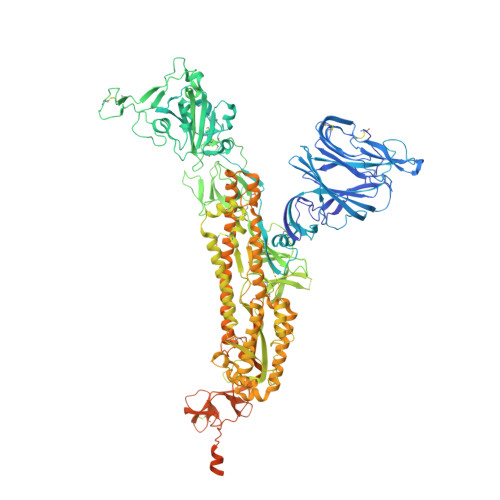
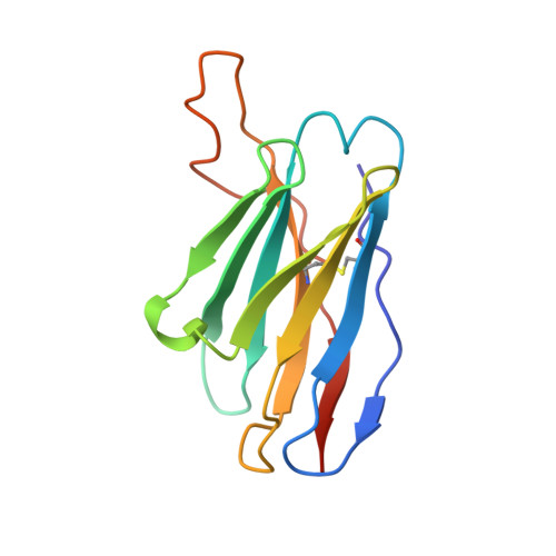
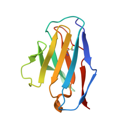
    As the coronavirus disease 2019 (COVID-19) pandemic continues, there is a strong need for highly potent monoclonal antibodies (mAbs) that are resistant against severe acute respiratory syndrome coronavirus 2 (SARS-CoV-2) variants of concern (VoCs). Here, we evaluate the potency of the previously described mAb J08 against these variants using cell-based assays and delve into the molecular details of the binding interaction using cryoelectron microscopy (cryo-EM) and X-ray crystallography. We show that mAb J08 has low nanomolar affinity against most VoCs and binds high on the receptor binding domain (RBD) ridge, away from many VoC mutations. These findings further validate the phase II/III human clinical trial underway using mAb J08 as a monoclonal therapy.


  • Organizational Affiliation
    • Department of Integrative Structural and Computational Biology, The Scripps Research Institute, La Jolla, CA 92037.

Macromolecule Content 

  • Total Structure Weight: 506.38 kDa 
  • Atom Count: 30,795 
  • Modeled Residue Count: 3,894 
  • Deposited Residue Count: 4,533 
  • Unique protein chains: 3

Macromolecules

Find similar proteins by:|  3D Structure
Entity ID: 1
MoleculeChains  Sequence LengthOrganismDetailsImage
Spike glycoproteinA,
D [auth B],
G [auth E]
1,280Severe acute respiratory syndrome coronavirus 2Mutation(s): 11 
Gene Names: S2
UniProt
Find proteins for P0DTC2 (Severe acute respiratory syndrome coronavirus 2)
Explore P0DTC2 
Go to UniProtKB:  P0DTC2
Entity Groups
Sequence Clusters30% Identity50% Identity70% Identity90% Identity95% Identity100% Identity
UniProt GroupP0DTC2
Glycosylation
Glycosylation Sites: 8Go to GlyGen: P0DTC2-1
Sequence Annotations
Expand
Reference Sequence
Find similar proteins by:|  3D Structure
Entity ID: 2
MoleculeChains  Sequence LengthOrganismDetailsImage
J08 fragment antigen binding heavy chain variable domainB [auth H],
E [auth C],
H [auth F]
126Homo sapiensMutation(s): 0 
Find similar proteins by:|  3D Structure
Entity ID: 3
MoleculeChains  Sequence LengthOrganismDetailsImage
J08 fragment antigen binding light chain variable domainC [auth L],
F [auth D],
I [auth G]
105Homo sapiensMutation(s): 0 

Oligosaccharides

Help  
Entity ID: 4
MoleculeChains Length2D Diagram GlycosylationD Interactions
2-acetamido-2-deoxy-beta-D-glucopyranose-(1-4)-2-acetamido-2-deoxy-beta-D-glucopyranose
J [auth I],
K [auth J],
L [auth K],
M,
N,
J [auth I],
K [auth J],
L [auth K],
M,
N,
O,
P,
Q,
R
2N-Glycosylation
Glycosylation Resources
GlyTouCan: G42666HT
GlyCosmos: G42666HT
GlyGen: G42666HT

Small Molecules

Ligands 1 Unique
IDChains Name / Formula / InChI Key2D Diagram3D Interactions
NAG

Query on NAG


Download:Ideal Coordinates CCD File
AA [auth B]
BA [auth B]
CA [auth E]
DA [auth E]
EA [auth E]
AA [auth B],
BA [auth B],
CA [auth E],
DA [auth E],
EA [auth E],
FA [auth E],
GA [auth E],
S [auth A],
T [auth A],
U [auth A],
V [auth A],
W [auth A],
X [auth B],
Y [auth B],
Z [auth B]
2-acetamido-2-deoxy-beta-D-glucopyranose
C8 H15 N O6
OVRNDRQMDRJTHS-FMDGEEDCSA-N

Experimental Data & Validation

Experimental Data

  • Method: ELECTRON MICROSCOPY
  • Resolution: 3.40 Å
  • Aggregation State: PARTICLE 
  • Reconstruction Method: SINGLE PARTICLE 
EM Software:
TaskSoftware PackageVersion
MODEL REFINEMENTRosetta
RECONSTRUCTIONcryoSPARC3.2
MODEL REFINEMENTPHENIX

Structure Validation

View Full Validation Report


Entry History 

& Funding Information

Deposition Data


Funding OrganizationLocationGrant Number
Bill & Melinda Gates FoundationItalyOPP1170236
Bill & Melinda Gates FoundationItalyINV-004923

Revision History  (Full details and data files)

  • Version 1.0: 2022-05-11
    Type: Initial release
  • Version 1.1: 2022-05-25
    Changes: Database references
  • Version 1.2: 2024-11-13
    Changes: Data collection, Structure summary
  • Version 1.3: 2025-05-28
    Changes: Data collection