7S2K

Crystal structure of sulfonamide resistance enzyme Sul2 in complex with 7,8-dihydropteroate, magnesium, and pyrophosphate


Experimental Data Snapshot

  • Method: X-RAY DIFFRACTION
  • Resolution: 1.74 Å
  • R-Value Free: 0.213 
  • R-Value Work: 0.179 
  • R-Value Observed: 0.180 

wwPDB Validation   3D Report Full Report


Ligand Structure Quality Assessment 


This is version 1.2 of the entry. See complete history


Literature

Molecular mechanism of plasmid-borne resistance to sulfonamide antibiotics.

Venkatesan, M.Fruci, M.Verellen, L.A.Skarina, T.Mesa, N.Flick, R.Pham, C.Mahadevan, R.Stogios, P.J.Savchenko, A.

(2023) Nat Commun 14: 4031-4031

  • DOI: https://doi.org/10.1038/s41467-023-39778-7
  • Primary Citation of Related Structures:  
    7S2I, 7S2J, 7S2K, 7S2L, 7S2M, 7TQ1, 8SCD

  • PubMed Abstract: 

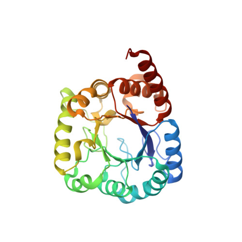
    The sulfonamides (sulfas) are the oldest class of antibacterial drugs and inhibit the bacterial dihydropteroate synthase (DHPS, encoded by folP), through chemical mimicry of its co-substrate p-aminobenzoic acid (pABA). Resistance to sulfa drugs is mediated either by mutations in folP or acquisition of sul genes, which code for sulfa-insensitive, divergent DHPS enzymes. While the molecular basis of resistance through folP mutations is well understood, the mechanisms mediating sul-based resistance have not been investigated in detail. Here, we determine crystal structures of the most common Sul enzyme types (Sul1, Sul2 and Sul3) in multiple ligand-bound states, revealing a substantial reorganization of their pABA-interaction region relative to the corresponding region of DHPS. We use biochemical and biophysical assays, mutational analysis, and in trans complementation of E. coli ΔfolP to show that a Phe-Gly sequence enables the Sul enzymes to discriminate against sulfas while retaining pABA binding and is necessary for broad resistance to sulfonamides. Experimental evolution of E. coli results in a strain harboring a sulfa-resistant DHPS variant that carries a Phe-Gly insertion in its active site, recapitulating this molecular mechanism. We also show that Sul enzymes possess increased active site conformational dynamics relative to DHPS, which could contribute to substrate discrimination. Our results reveal the molecular foundation for Sul-mediated drug resistance and facilitate the potential development of new sulfas less prone to resistance.


  • Organizational Affiliation

    Department of Chemical Engineering and Applied Chemistry, University of Toronto, Toronto, ON, M5S 1A4, Canada.


Macromolecules
Find similar proteins by:  (by identity cutoff)  |  3D Structure
Entity ID: 1
MoleculeChains Sequence LengthOrganismDetailsImage
Sul2
A, B
271uncultured bacteriumMutation(s): 0 
UniProt
Find proteins for P0AC11 (Escherichia coli)
Explore P0AC11 
Go to UniProtKB:  P0AC11
Entity Groups  
Sequence Clusters30% Identity50% Identity70% Identity90% Identity95% Identity100% Identity
UniProt GroupP0AC11
Sequence Annotations
Expand
  • Reference Sequence
Small Molecules
Ligands 6 Unique
IDChains Name / Formula / InChI Key2D Diagram3D Interactions
78H (Subject of Investigation/LOI)
Query on 78H

Download Ideal Coordinates CCD File 
C [auth A],
H [auth B]
7,8-DIHYDROPTEROATE
C14 H14 N6 O3
WBFYVDCHGVNRBH-UHFFFAOYSA-N
POP (Subject of Investigation/LOI)
Query on POP

Download Ideal Coordinates CCD File 
D [auth A]PYROPHOSPHATE 2-
H2 O7 P2
XPPKVPWEQAFLFU-UHFFFAOYSA-L
PAB (Subject of Investigation/LOI)
Query on PAB

Download Ideal Coordinates CCD File 
I [auth B]4-AMINOBENZOIC ACID
C7 H7 N O2
ALYNCZNDIQEVRV-UHFFFAOYSA-N
GOL
Query on GOL

Download Ideal Coordinates CCD File 
F [auth A]GLYCEROL
C3 H8 O3
PEDCQBHIVMGVHV-UHFFFAOYSA-N
CL
Query on CL

Download Ideal Coordinates CCD File 
G [auth A]CHLORIDE ION
Cl
VEXZGXHMUGYJMC-UHFFFAOYSA-M
MG (Subject of Investigation/LOI)
Query on MG

Download Ideal Coordinates CCD File 
E [auth A]MAGNESIUM ION
Mg
JLVVSXFLKOJNIY-UHFFFAOYSA-N
Experimental Data & Validation

Experimental Data

  • Method: X-RAY DIFFRACTION
  • Resolution: 1.74 Å
  • R-Value Free: 0.213 
  • R-Value Work: 0.179 
  • R-Value Observed: 0.180 
  • Space Group: P 1 21 1
Unit Cell:
Length ( Å )Angle ( ˚ )
a = 45.73α = 90
b = 74.243β = 96.075
c = 67.989γ = 90
Software Package:
Software NamePurpose
PHENIXrefinement
HKL-3000data reduction
HKL-3000data scaling
PHENIXphasing
PHENIXmodel building
Cootmodel building

Structure Validation

View Full Validation Report


Ligand Structure Quality Assessment 


Entry History & Funding Information

Deposition Data


Funding OrganizationLocationGrant Number
National Institutes of Health/National Institute Of Allergy and Infectious Diseases (NIH/NIAID)United StatesHHSN272201700060C

Revision History  (Full details and data files)

  • Version 1.0: 2023-05-03
    Type: Initial release
  • Version 1.1: 2023-07-19
    Changes: Database references
  • Version 1.2: 2023-10-25
    Changes: Data collection, Refinement description