7R86

Structure of mouse BAI1 (ADGRB1) in complex with mouse Nogo receptor (RTN4R)


Experimental Data Snapshot

  • Method: X-RAY DIFFRACTION
  • Resolution: 1.65 Å
  • R-Value Free: 0.195 
  • R-Value Work: 0.165 
  • R-Value Observed: 0.166 

wwPDB Validation   3D Report Full Report


Ligand Structure Quality Assessment 


This is version 1.2 of the entry. See complete history


Literature

RTN4/NoGo-receptor binding to BAI adhesion-GPCRs regulates neuronal development.

Wang, J.Miao, Y.Wicklein, R.Sun, Z.Wang, J.Jude, K.M.Fernandes, R.A.Merrill, S.A.Wernig, M.Garcia, K.C.Sudhof, T.C.

(2021) Cell 184: 5869-5885.e25

  • DOI: https://doi.org/10.1016/j.cell.2021.10.016
  • Primary Citation of Related Structures:  
    7R84, 7R85, 7R86

  • PubMed Abstract: 

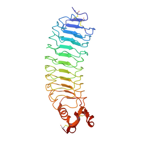
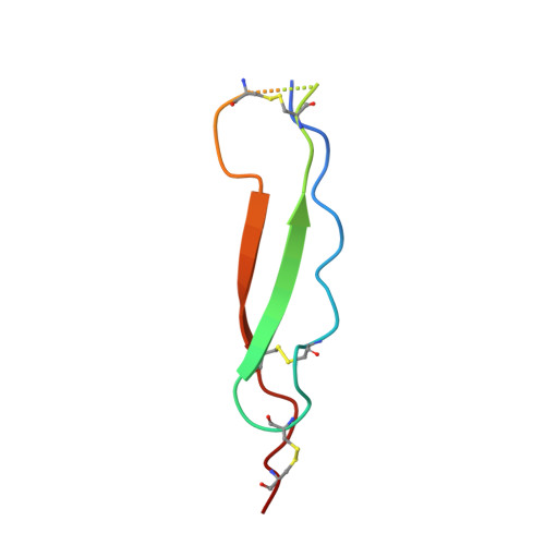
    RTN4-binding proteins were widely studied as "NoGo" receptors, but their physiological interactors and roles remain elusive. Similarly, BAI adhesion-GPCRs were associated with numerous activities, but their ligands and functions remain unclear. Using unbiased approaches, we observed an unexpected convergence: RTN4 receptors are high-affinity ligands for BAI adhesion-GPCRs. A single thrombospondin type 1-repeat (TSR) domain of BAIs binds to the leucine-rich repeat domain of all three RTN4-receptor isoforms with nanomolar affinity. In the 1.65 Å crystal structure of the BAI1/RTN4-receptor complex, C-mannosylation of tryptophan and O-fucosylation of threonine in the BAI TSR-domains creates a RTN4-receptor/BAI interface shaped by unusual glycoconjugates that enables high-affinity interactions. In human neurons, RTN4 receptors regulate dendritic arborization, axonal elongation, and synapse formation by differential binding to glial versus neuronal BAIs, thereby controlling neural network activity. Thus, BAI binding to RTN4/NoGo receptors represents a receptor-ligand axis that, enabled by rare post-translational modifications, controls development of synaptic circuits.


  • Organizational Affiliation

    Department of Molecular and Cellular Physiology, Stanford University School of Medicine, Stanford, CA 94305, USA.


Macromolecules
Find similar proteins by:  (by identity cutoff)  |  3D Structure
Entity ID: 1
MoleculeChains Sequence LengthOrganismDetailsImage
Reticulon-4 receptor
A, B
285Mus musculusMutation(s): 0 
Gene Names: Rtn4rNgr1Nogor
UniProt
Find proteins for Q99PI8 (Mus musculus)
Explore Q99PI8 
Go to UniProtKB:  Q99PI8
Entity Groups  
Sequence Clusters30% Identity50% Identity70% Identity90% Identity95% Identity100% Identity
UniProt GroupQ99PI8
Sequence Annotations
Expand
  • Reference Sequence
Find similar proteins by:  (by identity cutoff)  |  3D Structure
Entity ID: 2
MoleculeChains Sequence LengthOrganismDetailsImage
Vasculostatin-120
C, D
54Mus musculusMutation(s): 0 
Gene Names: Adgrb1Bai1
UniProt & NIH Common Fund Data Resources
Find proteins for Q3UHD1 (Mus musculus)
Explore Q3UHD1 
Go to UniProtKB:  Q3UHD1
IMPC:  MGI:1933736
Entity Groups  
Sequence Clusters30% Identity50% Identity70% Identity90% Identity95% Identity100% Identity
UniProt GroupQ3UHD1
Sequence Annotations
Expand
  • Reference Sequence
Oligosaccharides

Help

Entity ID: 3
MoleculeChains Length2D Diagram Glycosylation3D Interactions
beta-D-glucopyranose-(1-3)-alpha-L-fucopyranose
E, F
2O-Glycosylation
Glycosylation Resources
GlyTouCan:  G36855WW
GlyCosmos:  G36855WW
GlyGen:  G36855WW
Small Molecules
Ligands 7 Unique
IDChains Name / Formula / InChI Key2D Diagram3D Interactions
NAG
Query on NAG

Download Ideal Coordinates CCD File 
G [auth A],
H [auth A],
Q [auth B],
R [auth B]
2-acetamido-2-deoxy-beta-D-glucopyranose
C8 H15 N O6
OVRNDRQMDRJTHS-FMDGEEDCSA-N
PG4
Query on PG4

Download Ideal Coordinates CCD File 
J [auth A]TETRAETHYLENE GLYCOL
C8 H18 O5
UWHCKJMYHZGTIT-UHFFFAOYSA-N
MAN
Query on MAN

Download Ideal Coordinates CCD File 
AA [auth D],
W [auth C],
X [auth C],
Z [auth D]
alpha-D-mannopyranose
C6 H12 O6
WQZGKKKJIJFFOK-PQMKYFCFSA-N
PAB
Query on PAB

Download Ideal Coordinates CCD File 
I [auth A]4-AMINOBENZOIC ACID
C7 H7 N O2
ALYNCZNDIQEVRV-UHFFFAOYSA-N
EDO
Query on EDO

Download Ideal Coordinates CCD File 
K [auth A]
L [auth A]
M [auth A]
N [auth A]
O [auth A]
K [auth A],
L [auth A],
M [auth A],
N [auth A],
O [auth A],
S [auth B],
T [auth B],
Y [auth C]
1,2-ETHANEDIOL
C2 H6 O2
LYCAIKOWRPUZTN-UHFFFAOYSA-N
ACT
Query on ACT

Download Ideal Coordinates CCD File 
U [auth B]ACETATE ION
C2 H3 O2
QTBSBXVTEAMEQO-UHFFFAOYSA-M
CL
Query on CL

Download Ideal Coordinates CCD File 
P [auth A],
V [auth B]
CHLORIDE ION
Cl
VEXZGXHMUGYJMC-UHFFFAOYSA-M
Experimental Data & Validation

Experimental Data

Unit Cell:
Length ( Å )Angle ( ˚ )
a = 63.468α = 90
b = 95.315β = 105.39
c = 69.682γ = 90
Software Package:
Software NamePurpose
Aimlessdata scaling
PHENIXrefinement
PDB_EXTRACTdata extraction
XDSdata reduction
PHASERphasing

Structure Validation

View Full Validation Report


Ligand Structure Quality Assessment 


Entry History & Funding Information

Deposition Data


Funding OrganizationLocationGrant Number
Howard Hughes Medical Institute (HHMI)United States--

Revision History  (Full details and data files)

  • Version 1.0: 2021-11-10
    Type: Initial release
  • Version 1.1: 2022-05-25
    Changes: Database references
  • Version 1.2: 2023-10-18
    Changes: Data collection, Refinement description