7QPI

Structure of lamprey VDR in complex with 1,25D3


Experimental Data Snapshot

  • Method: X-RAY DIFFRACTION
  • Resolution: 2.50 Å
  • R-Value Free: 0.256 
  • R-Value Work: 0.182 
  • R-Value Observed: 0.186 

wwPDB Validation   3D Report Full Report


Ligand Structure Quality Assessment 


This is version 1.1 of the entry. See complete history


Literature

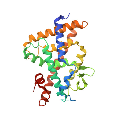
Advances in Vitamin D Receptor Function and Evolution Based on the 3D Structure of the Lamprey Ligand-Binding Domain.

Sigueiro, R.Bianchetti, L.Peluso-Iltis, C.Chalhoub, S.Dejaegere, A.Osz, J.Rochel, N.

(2022) J Med Chem 65: 5821-5829

  • DOI: https://doi.org/10.1021/acs.jmedchem.2c00171
  • Primary Citation of Related Structures:  
    7QPI, 7QPP

  • PubMed Abstract: 

    1α,25-dihydroxyvitamin D3 (1,25D 3 ) regulates many physiological processes in vertebrates by binding to the vitamin D receptor (VDR). Phylogenetic analysis indicates that jawless fishes are the most basal vertebrates exhibiting a VDR gene. To elucidate the mechanism driving VDR activation during evolution, we determined the crystal structure of the VDR ligand-binding domain (LBD) complex from the basal vertebrate Petromyzon marinus , sea lamprey (lVDR). Comparison of three-dimensional crystal structures of the lVDR-1,25D 3 complex with higher vertebrate VDR-1,25D 3 structures suggests that 1,25D 3 binds to lVDR similarly to human VDR, but with unique features for lVDR around linker regions between H11 and H12 and between H9 and H10. These structural differences may contribute to the marked species differences in transcriptional responses. Furthermore, residue co-evolution analysis of VDR across vertebrates identifies amino acid positions in H9 and the large insertion domain VDR LBD specific as correlated.


  • Organizational Affiliation

    Institut de Génétique et de Biologie Moléculaire et Cellulaire (IGBMC), 67400 Illkirch, France.


Macromolecules
Find similar proteins by:  (by identity cutoff)  |  3D Structure
Entity ID: 1
MoleculeChains Sequence LengthOrganismDetailsImage
Vitamin D receptor287Petromyzon marinusMutation(s): 0 
Gene Names: VDR
UniProt
Find proteins for S4R4S2 (Petromyzon marinus)
Explore S4R4S2 
Go to UniProtKB:  S4R4S2
Entity Groups  
Sequence Clusters30% Identity50% Identity70% Identity90% Identity95% Identity100% Identity
UniProt GroupS4R4S2
Sequence Annotations
Expand
  • Reference Sequence

Find similar proteins by:  Sequence   |   3D Structure  

Entity ID: 2
MoleculeChains Sequence LengthOrganismDetailsImage
Nuclear receptor coactivator 1B [auth U]13Homo sapiensMutation(s): 0 
EC: 2.3.1.48
UniProt & NIH Common Fund Data Resources
Find proteins for Q15788 (Homo sapiens)
Explore Q15788 
Go to UniProtKB:  Q15788
PHAROS:  Q15788
GTEx:  ENSG00000084676 
Entity Groups  
Sequence Clusters30% Identity50% Identity70% Identity90% Identity95% Identity100% Identity
UniProt GroupQ15788
Sequence Annotations
Expand
  • Reference Sequence
Small Molecules
Ligands 2 Unique
IDChains Name / Formula / InChI Key2D Diagram3D Interactions
VDX (Subject of Investigation/LOI)
Query on VDX

Download Ideal Coordinates CCD File 
C [auth A]5-{2-[1-(5-HYDROXY-1,5-DIMETHYL-HEXYL)-7A-METHYL-OCTAHYDRO-INDEN-4-YLIDENE]-ETHYLIDENE}-4-METHYLENE-CYCLOHEXANE-1,3-DIOL
C27 H44 O3
GMRQFYUYWCNGIN-NKMMMXOESA-N
MG
Query on MG

Download Ideal Coordinates CCD File 
D [auth A],
E [auth A]
MAGNESIUM ION
Mg
JLVVSXFLKOJNIY-UHFFFAOYSA-N
Experimental Data & Validation

Experimental Data

  • Method: X-RAY DIFFRACTION
  • Resolution: 2.50 Å
  • R-Value Free: 0.256 
  • R-Value Work: 0.182 
  • R-Value Observed: 0.186 
  • Space Group: P 21 21 21
Unit Cell:
Length ( Å )Angle ( ˚ )
a = 37.32α = 90
b = 66.17β = 90
c = 105.69γ = 90
Software Package:
Software NamePurpose
PHENIXrefinement
PDB_EXTRACTdata extraction
XDSdata reduction
XDSdata scaling
PHENIXphasing

Structure Validation

View Full Validation Report


Ligand Structure Quality Assessment 


Entry History & Funding Information

Deposition Data

  • Released Date: 2022-12-14 
  • Deposition Author(s): Rochel, N.

Funding OrganizationLocationGrant Number
Agence Nationale de la Recherche (ANR)France--

Revision History  (Full details and data files)

  • Version 1.0: 2022-12-14
    Type: Initial release
  • Version 1.1: 2024-01-31
    Changes: Data collection, Refinement description