7PX4

Crystal structure of the adenosine A2A receptor (A2A-PSB1-bRIL) in complex with preladenant conjugate PSB-2113


Experimental Data Snapshot

  • Method: X-RAY DIFFRACTION
  • Resolution: 2.25 Å
  • R-Value Free: 0.240 
  • R-Value Work: 0.196 
  • R-Value Observed: 0.198 

wwPDB Validation   3D Report Full Report


Ligand Structure Quality Assessment 


This is version 1.3 of the entry. See complete history


Literature

Single Stabilizing Point Mutation Enables High-Resolution Co-Crystal Structures of the Adenosine A 2A Receptor with Preladenant Conjugates.

Claff, T.Klapschinski, T.A.Tiruttani Subhramanyam, U.K.Vaassen, V.J.Schlegel, J.G.Vielmuth, C.Voss, J.H.Labahn, J.Muller, C.E.

(2022) Angew Chem Int Ed Engl 61: e202115545-e202115545

  • DOI: https://doi.org/10.1002/anie.202115545
  • Primary Citation of Related Structures:  
    7PX4, 7PYR

  • PubMed Abstract: 

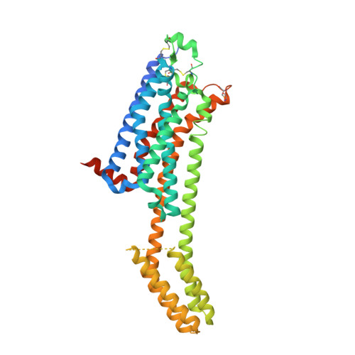
    The G protein-coupled adenosine A 2A receptor (A 2A AR) is an important new (potential) drug target in immuno-oncology, and for neurodegenerative diseases. Preladenant and its derivatives belong to the most potent A 2A AR antagonists displaying exceptional selectivity. While crystal structures of the human A 2A AR have been solved, mostly using the A 2A -StaR2 protein that bears 9 point mutations, co-crystallization with Preladenant derivatives has so far been elusive. We developed a new A 2A AR construct harboring a single point mutation (S91 3.39 K) which renders it extremely thermostable. This allowed the co-crystallization of two novel Preladenant derivatives, the polyethylene glycol-conjugated (PEGylated) PSB-2113, and the fluorophore-labeled PSB-2115. The obtained crystal structures (2.25 Å and 2.6 Å resolution) provide explanations for the high potency and selectivity of Preladenant derivatives. They represent the first crystal structures of a GPCR in complex with PEG- and fluorophore-conjugated ligands. The applied strategy is predicted to be applicable to further class A GPCRs.


  • Organizational Affiliation

    Pharmaceutical Institute, Pharmaceutical & Medicinal Chemistry, University of Bonn, An der Immenburg 4, 53121, Bonn, Germany.


Macromolecules
Find similar proteins by:  (by identity cutoff)  |  3D Structure
Entity ID: 1
MoleculeChains Sequence LengthOrganismDetailsImage
Adenosine receptor A2a,Soluble cytochrome b562,Adenosine receptor A2a447Homo sapiensEscherichia coliMutation(s): 4 
Gene Names: ADORA2AADORA2cybC
Membrane Entity: Yes 
UniProt & NIH Common Fund Data Resources
Find proteins for P0ABE7 (Escherichia coli)
Explore P0ABE7 
Go to UniProtKB:  P0ABE7
Find proteins for P29274 (Homo sapiens)
Explore P29274 
Go to UniProtKB:  P29274
PHAROS:  P29274
GTEx:  ENSG00000128271 
Entity Groups  
Sequence Clusters30% Identity50% Identity70% Identity90% Identity95% Identity100% Identity
UniProt GroupsP0ABE7P29274
Sequence Annotations
Expand
  • Reference Sequence
Small Molecules
Ligands 5 Unique
IDChains Name / Formula / InChI Key2D Diagram3D Interactions
8E2 (Subject of Investigation/LOI)
Query on 8E2

Download Ideal Coordinates CCD File 
E [auth A]Preladenant conjugate PSB-2113
C38 H52 N10 O9
BIWDHFUPTSCUTC-UHFFFAOYSA-N
CLR
Query on CLR

Download Ideal Coordinates CCD File 
B [auth A],
C [auth A],
D [auth A]
CHOLESTEROL
C27 H46 O
HVYWMOMLDIMFJA-DPAQBDIFSA-N
OLC
Query on OLC

Download Ideal Coordinates CCD File 
FA [auth A],
GA [auth A],
HA [auth A]
(2R)-2,3-dihydroxypropyl (9Z)-octadec-9-enoate
C21 H40 O4
RZRNAYUHWVFMIP-GDCKJWNLSA-N
OLA
Query on OLA

Download Ideal Coordinates CCD File 
AA [auth A]
BA [auth A]
CA [auth A]
DA [auth A]
EA [auth A]
AA [auth A],
BA [auth A],
CA [auth A],
DA [auth A],
EA [auth A],
F [auth A],
G [auth A],
H [auth A],
I [auth A],
J [auth A],
K [auth A],
L [auth A],
M [auth A],
N [auth A],
O [auth A],
P [auth A],
Q [auth A],
R [auth A],
S [auth A],
T [auth A],
U [auth A],
V [auth A],
W [auth A],
X [auth A],
Y [auth A],
Z [auth A]
OLEIC ACID
C18 H34 O2
ZQPPMHVWECSIRJ-KTKRTIGZSA-N
PEG
Query on PEG

Download Ideal Coordinates CCD File 
IA [auth A],
JA [auth A]
DI(HYDROXYETHYL)ETHER
C4 H10 O3
MTHSVFCYNBDYFN-UHFFFAOYSA-N
Experimental Data & Validation

Experimental Data

  • Method: X-RAY DIFFRACTION
  • Resolution: 2.25 Å
  • R-Value Free: 0.240 
  • R-Value Work: 0.196 
  • R-Value Observed: 0.198 
  • Space Group: C 2 2 21
Unit Cell:
Length ( Å )Angle ( ˚ )
a = 39.6α = 90
b = 180.262β = 90
c = 139.483γ = 90
Software Package:
Software NamePurpose
PHENIXrefinement
XDSdata reduction
XDSdata scaling
PHASERphasing
Cootmodel building

Structure Validation

View Full Validation Report


Ligand Structure Quality Assessment 


Entry History & Funding Information

Deposition Data


Funding OrganizationLocationGrant Number
German Research Foundation (DFG)GermanySFB1328
German Research Foundation (DFG)GermanyFOR2327
German Federal Ministry for Education and ResearchGermany--

Revision History  (Full details and data files)

  • Version 1.0: 2022-03-02
    Type: Initial release
  • Version 1.1: 2022-04-06
    Changes: Database references
  • Version 1.2: 2022-05-25
    Changes: Database references
  • Version 1.3: 2024-01-31
    Changes: Data collection, Refinement description