7PLS

Cryo-EM structures of human fucosidase FucA1 reveal insight into substate recognition and catalysis.


Experimental Data Snapshot

  • Method: ELECTRON MICROSCOPY
  • Resolution: 2.49 Å
  • Aggregation State: PARTICLE 
  • Reconstruction Method: SINGLE PARTICLE 

wwPDB Validation   3D Report Full Report


This is version 1.1 of the entry. See complete history


Literature

Cryo-EM structures of human fucosidase FucA1 reveal insight into substrate recognition and catalysis.

Armstrong, Z.Meek, R.W.Wu, L.Blaza, J.N.Davies, G.J.

(2022) Structure 30: 1443

  • DOI: https://doi.org/10.1016/j.str.2022.07.001
  • Primary Citation of Related Structures:  
    7PLS, 7PM4

  • PubMed Abstract: 

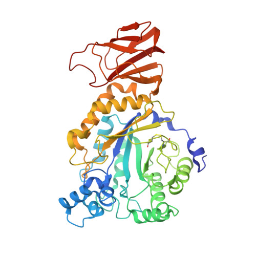
    Enzymatic hydrolysis of α-L-fucose from fucosylated glycoconjugates is consequential in bacterial infections and the neurodegenerative lysosomal storage disorder fucosidosis. Understanding human α-L-fucosidase catalysis, in an effort toward drug design, has been hindered by the absence of three-dimensional structural data for any animal fucosidase. Here, we have used cryoelectron microscopy (cryo-EM) to determine the structure of human lysosomal α-L-fucosidase (FucA1) in both an unliganded state and in complex with the inhibitor deoxyfuconojirimycin. These structures, determined at 2.49 Å resolution, reveal the homotetrameric structure of FucA1, the architecture of the catalytic center, and the location of both natural population variations and disease-causing mutations. Furthermore, this work has conclusively identified the hitherto contentious identity of the catalytic acid/base as aspartate-276, representing a shift from both the canonical glutamate acid/base residue and a previously proposed glutamate residue. These findings have furthered our understanding of how FucA1 functions in both health and disease.


  • Organizational Affiliation

    Department of Chemistry, Structural Biology Laboratory, University of York, Heslington, York YO10 5DD, UK.


Macromolecules
Find similar proteins by:  (by identity cutoff)  |  3D Structure
Entity ID: 1
MoleculeChains Sequence LengthOrganismDetailsImage
Tissue alpha-L-fucosidase
A, B, C, D
436Homo sapiensMutation(s): 0 
Gene Names: FUCA1Nbla10230
EC: 3.2.1.51
UniProt & NIH Common Fund Data Resources
Find proteins for P04066 (Homo sapiens)
Explore P04066 
Go to UniProtKB:  P04066
PHAROS:  P04066
GTEx:  ENSG00000179163 
Entity Groups  
Sequence Clusters30% Identity50% Identity70% Identity90% Identity95% Identity100% Identity
UniProt GroupP04066
Sequence Annotations
Expand
  • Reference Sequence
Experimental Data & Validation

Experimental Data

  • Method: ELECTRON MICROSCOPY
  • Resolution: 2.49 Å
  • Aggregation State: PARTICLE 
  • Reconstruction Method: SINGLE PARTICLE 
EM Software:
TaskSoftware PackageVersion
RECONSTRUCTIONRELION3.1.2
MODEL REFINEMENTPHENIX1.19

Structure Validation

View Full Validation Report


Entry History & Funding Information

Deposition Data


Funding OrganizationLocationGrant Number
Royal SocietyUnited Kingdom180016
Biotechnology and Biological Sciences Research Council (BBSRC)United KingdomR001162
UK Research and Innovation (UKRI)United KingdomT040742
Wellcome TrustUnited Kingdom218579

Revision History  (Full details and data files)

  • Version 1.0: 2022-08-10
    Type: Initial release
  • Version 1.1: 2022-10-19
    Changes: Database references