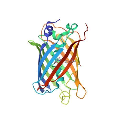
Structure and Function of Redox-Sensitive Superfolder Green Fluorescent Protein Variant.
Heimsch, K.C., Gertzen, C.G.W., Schuh, A.K., Nietzel, T., Rahlfs, S., Przyborski, J.M., Gohlke, H., Schwarzlander, M., Becker, K., Fritz-Wolf, K.(2022) Antioxid Redox Signal 37: 1-18
- PubMed: 35072524
- DOI: https://doi.org/10.1089/ars.2021.0234
- Primary Citation of Related Structures:
7PCA, 7PCZ, 7PD0 - PubMed Abstract:
Aims: Genetically encoded green fluorescent protein (GFP)-based redox biosensors are widely used to monitor specific and dynamic redox processes in living cells. Over the last few years, various biosensors for a variety of applications were engineered and enhanced to match the organism and cellular environments, which should be investigated. In this context, the unicellular intraerythrocytic parasite Plasmodium , the causative agent of malaria, represents a challenge, as the small size of the organism results in weak fluorescence signals that complicate precise measurements, especially for cell compartment-specific observations. To address this, we have functionally and structurally characterized an enhanced redox biosensor superfolder roGFP2 (sfroGFP2). Results: SfroGFP2 retains roGFP2-like behavior, yet with improved fluorescence intensity (FI) in cellulo . SfroGFP2-based redox biosensors are pH insensitive in a physiological pH range and show midpoint potentials comparable with roGFP2-based redox biosensors. Using crystallography and rigidity theory, we identified the superfolding mutations as being responsible for improved structural stability of the biosensor in a redox-sensitive environment, thus explaining the improved FI in cellulo . Innovation: This work provides insight into the structure and function of GFP-based redox biosensors. It describes an improved redox biosensor (sfroGFP2) suitable for measuring oxidizing effects within small cells where applicability of other redox sensor variants is limited. Conclusion: Improved structural stability of sfroGFP2 gives rise to increased FI in cellulo . Fusion to hGrx1 (human glutaredoxin-1) provides the hitherto most suitable biosensor for measuring oxidizing effects in Plasmodium . This sensor is of major interest for studying glutathione redox changes in small cells, as well as subcellular compartments in general. Antioxid. Redox Signal. 37, 1-18.
- Biochemistry and Molecular Biology, Interdisciplinary Research Center, Justus Liebig University Giessen, Giessen, Germany.
Organizational Affiliation:


