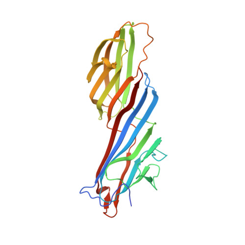
7OFP

Apo Structure of Mu2 Adaptin Subunit (Ap50) Of AP2 Clathrin Adaptor


Experimental Data Snapshot

  • Method: X-RAY DIFFRACTION
  • Resolution: 1.92 Å
  • R-Value Free: 0.210 
  • R-Value Work: 0.188 
  • R-Value Observed: 0.189 

wwPDB Validation   3D Report Full Report


This is version 1.1 of the entry. See complete history


Literature

FCHO controls AP2's initiating role in endocytosis through a PtdIns(4,5)P 2 -dependent switch.

Zaccai, N.R.Kadlecova, Z.Dickson, V.K.Korobchevskaya, K.Kamenicky, J.Kovtun, O.Umasankar, P.K.Wrobel, A.G.Kaufman, J.G.G.Gray, S.R.Qu, K.Evans, P.R.Fritzsche, M.Sroubek, F.Honing, S.Briggs, J.A.G.Kelly, B.T.Owen, D.J.Traub, L.M.

(2022) Sci Adv 8: eabn2018-eabn2018

  • DOI: https://doi.org/10.1126/sciadv.abn2018
  • Primary Citation of Related Structures:  
    7OFP, 7OG1, 7OHI, 7OHO, 7OHZ, 7OI5, 7OIQ, 7OIT, 7Z5C

  • PubMed Abstract: 

    Clathrin-mediated endocytosis (CME) is the main mechanism by which mammalian cells control their cell surface proteome. Proper operation of the pivotal CME cargo adaptor AP2 requires membrane-localized Fer/Cip4 homology domain-only proteins (FCHO). Here, live-cell enhanced total internal reflection fluorescence-structured illumination microscopy shows that FCHO marks sites of clathrin-coated pit (CCP) initiation, which mature into uniform-sized CCPs comprising a central patch of AP2 and clathrin corralled by an FCHO/Epidermal growth factor potential receptor substrate number 15 (Eps15) ring. We dissect the network of interactions between the FCHO interdomain linker and AP2, which concentrates, orients, tethers, and partially destabilizes closed AP2 at the plasma membrane. AP2's subsequent membrane deposition drives its opening, which triggers FCHO displacement through steric competition with phosphatidylinositol 4,5-bisphosphate, clathrin, cargo, and CME accessory factors. FCHO can now relocate toward a CCP's outer edge to engage and activate further AP2s to drive CCP growth/maturation.


  • Organizational Affiliation

    CIMR, University of Cambridge, Biomedical Campus, Hills Road, Cambridge CB2 0XY, UK.


Macromolecules
Find similar proteins by:  (by identity cutoff)  |  3D Structure
Entity ID: 1
MoleculeChains Sequence LengthOrganismDetailsImage
AP-2 complex subunit mu
A, B
285Rattus norvegicusMutation(s): 0 
Gene Names: Ap2m1
UniProt
Find proteins for P84092 (Rattus norvegicus)
Explore P84092 
Go to UniProtKB:  P84092
Entity Groups  
Sequence Clusters30% Identity50% Identity70% Identity90% Identity95% Identity100% Identity
UniProt GroupP84092
Sequence Annotations
Expand
  • Reference Sequence
Small Molecules
Ligands 2 Unique
IDChains Name / Formula / InChI Key2D Diagram3D Interactions
CIT
Query on CIT

Download Ideal Coordinates CCD File 
I [auth A]CITRIC ACID
C6 H8 O7
KRKNYBCHXYNGOX-UHFFFAOYSA-N
GOL
Query on GOL

Download Ideal Coordinates CCD File 
C [auth A]
D [auth A]
E [auth A]
F [auth A]
G [auth A]
C [auth A],
D [auth A],
E [auth A],
F [auth A],
G [auth A],
H [auth A],
J [auth B],
K [auth B],
L [auth B]
GLYCEROL
C3 H8 O3
PEDCQBHIVMGVHV-UHFFFAOYSA-N
Experimental Data & Validation

Experimental Data

  • Method: X-RAY DIFFRACTION
  • Resolution: 1.92 Å
  • R-Value Free: 0.210 
  • R-Value Work: 0.188 
  • R-Value Observed: 0.189 
  • Space Group: P 65
Unit Cell:
Length ( Å )Angle ( ˚ )
a = 123.83α = 90
b = 123.83β = 90
c = 112.668γ = 120
Software Package:
Software NamePurpose
REFMACrefinement
PHENIXrefinement
DIALSdata reduction
DIALSdata scaling
PHASERphasing
STARANISOdata processing
autoPROCdata processing

Structure Validation

View Full Validation Report


Entry History & Funding Information

Deposition Data


Funding OrganizationLocationGrant Number
Wellcome TrustUnited Kingdom207455/Z/17/Z

Revision History  (Full details and data files)

  • Version 1.0: 2022-06-01
    Type: Initial release
  • Version 1.1: 2024-01-31
    Changes: Data collection, Refinement description