Recruitment of phospholipase C gamma 1 to the non-structural membrane protein pK15 of Kaposi Sarcoma-associated herpesvirus promotes its Src-dependent phosphorylation.
Samarina, N., Ssebyatika, G., Tikla, T., Waldmann, J.Y., Abere, B., Nanna, V., Marasco, M., Carlomagno, T., Krey, T., Schulz, T.F.(2021) PLoS Pathog 17: e1009635-e1009635
- PubMed: 34143834 Search on PubMedSearch on PubMed Central
- DOI: https://doi.org/10.1371/journal.ppat.1009635
- Primary Citation Related Structures:
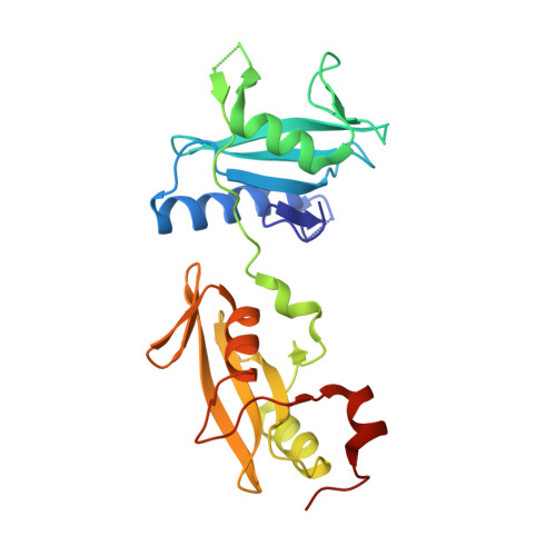
7NXE - PubMed Abstract:
Kaposi Sarcoma-associated herpesvirus (KSHV) causes three human malignancies, Kaposi Sarcoma (KS), Primary Effusion Lymphoma (PEL) and the plasma cell variant of multicentric Castleman's Disease (MCD), as well as an inflammatory cytokine syndrome (KICS). Its non-structural membrane protein, pK15, is among a limited set of viral proteins expressed in KSHV-infected KS tumor cells. Following its phosphorylation by Src family tyrosine kinases, pK15 recruits phospholipase C gamma 1 (PLCγ1) to activate downstream signaling cascades such as the MEK/ERK, NFkB and PI3K pathway, and thereby contributes to the increased proliferation and migration as well as the spindle cell morphology of KSHV-infected endothelial cells. Here, we show that a phosphorylated Y481EEVL motif in pK15 preferentially binds into the PLCγ1 C-terminal SH2 domain (cSH2), which is involved in conformational changes occurring during the activation of PLCγ1 by receptor tyrosine kinases. We determined the crystal structure of a pK15 12mer peptide containing the phosphorylated pK15 Y481EEVL motif in complex with a shortened PLCγ1 tandem SH2 (tSH2) domain. This structure demonstrates that the pK15 peptide binds to the PLCγ1 cSH2 domain in a position that is normally occupied by the linker region connecting the PLCγ1 cSH2 and SH3 domains. We also show that longer pK15 peptides containing the phosphorylated pK15 Y481EEVL motif can increase the Src-mediated phosphorylation of the PLCγ1 tSH2 region in vitro. This pK15-induced increase in Src-mediated phosphorylation of PLCγ1 can be inhibited with the small pK15-derived peptide which occupies the PLCγ1 cSH2 domain. Our findings thus suggest that pK15 may act as a scaffold protein to promote PLCγ1 activation in a manner similar to the cellular scaffold protein SLP-76, which has been shown to promote PLCγ1 activation in the context of T-cell receptor signaling. Reminiscent of its positional homologue in Epstein-Barr Virus, LMP2A, pK15 may therefore mimic aspects of antigen-receptor signaling. Our findings also suggest that it may be possible to inhibit the recruitment and activation of PLCγ1 pharmacologically.
- Institute of Virology, Hannover Medical School, Hannover, Germany.
Organizational Affiliation: