7MX4

CD1c with antigen analogue 1


Experimental Data Snapshot

  • Method: X-RAY DIFFRACTION
  • Resolution: 1.73 Å
  • R-Value Free: 0.239 
  • R-Value Work: 0.207 
  • R-Value Observed: 0.208 

wwPDB Validation   3D Report Full Report


Ligand Structure Quality Assessment 


This is version 1.1 of the entry. See complete history


Literature

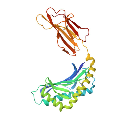
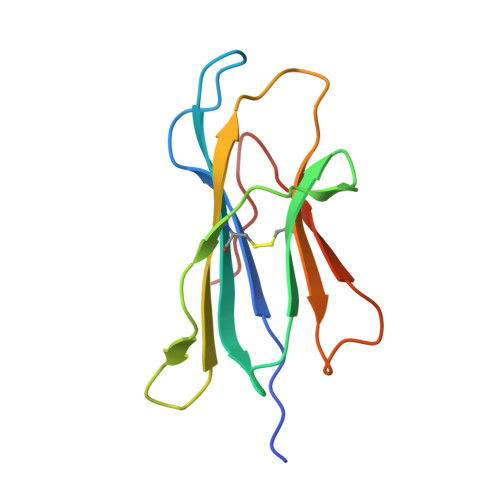
Rational design of a hydrolysis-resistant mycobacterial phosphoglycolipid antigen presented by CD1c to T cells.

Reijneveld, J.F.Marino, L.Cao, T.P.Cheng, T.Y.Dam, D.Shahine, A.Witte, M.D.Filippov, D.V.Suliman, S.van der Marel, G.A.Moody, D.B.Minnaard, A.J.Rossjohn, J.Codee, J.D.C.Van Rhijn, I.

(2021) J Biol Chem 297: 101197-101197

  • DOI: https://doi.org/10.1016/j.jbc.2021.101197
  • Primary Citation of Related Structures:  
    7MX4, 7MXF, 7MXH

  • PubMed Abstract: 

    Whereas proteolytic cleavage is crucial for peptide presentation by classical major histocompatibility complex (MHC) proteins to T cells, glycolipids presented by CD1 molecules are typically presented in an unmodified form. However, the mycobacterial lipid antigen mannosyl-β1-phosphomycoketide (MPM) may be processed through hydrolysis in antigen presenting cells, forming mannose and phosphomycoketide (PM). To further test the hypothesis that some lipid antigens are processed, and to generate antigens that lead to defined epitopes for future tuberculosis vaccines or diagnostic tests, we aimed to create hydrolysis-resistant MPM variants that retain their antigenicity. Here, we designed and tested three different, versatile synthetic strategies to chemically stabilize MPM analogs. Crystallographic studies of CD1c complexes with these three new MPM analogs showed anchoring of the lipid tail and phosphate group that is highly comparable to nature-identical MPM, with considerable conformational flexibility for the mannose head group. MPM-3, a difluoromethylene-modified version of MPM that is resistant to hydrolysis, showed altered recognition by cells, but not by CD1c proteins, supporting the cellular antigen processing hypothesis. Furthermore, the synthetic analogs elicited T cell responses that were cross-reactive with nature-identical MPM, fulfilling important requirements for future clinical use.


  • Organizational Affiliation

    Division of Rheumatology, Inflammation, and Immunity, Brigham and Women's Hospital and Harvard Medical School, Boston, Massachusetts, USA; Department of Infectious Diseases and Immunology, Faculty of Veterinary Medicine, Utrecht University, Utrecht, the Netherlands; Stratingh Institute for Chemistry, University of Groningen, Groningen, the Netherlands.


Macromolecules
Find similar proteins by:  (by identity cutoff)  |  3D Structure
Entity ID: 1
MoleculeChains Sequence LengthOrganismDetailsImage
T-cell surface glycoprotein CD1c/T-cell surface glycoprotein CD1b chimeric protein282Homo sapiensMutation(s): 5 
Gene Names: CD1CCD1B
UniProt & NIH Common Fund Data Resources
Find proteins for P29016 (Homo sapiens)
Explore P29016 
Go to UniProtKB:  P29016
PHAROS:  P29016
GTEx:  ENSG00000158485 
Find proteins for P29017 (Homo sapiens)
Explore P29017 
Go to UniProtKB:  P29017
PHAROS:  P29017
GTEx:  ENSG00000158481 
Entity Groups  
Sequence Clusters30% Identity50% Identity70% Identity90% Identity95% Identity100% Identity
UniProt GroupsP29017P29016
Sequence Annotations
Expand
  • Reference Sequence
Find similar proteins by:  (by identity cutoff)  |  3D Structure
Entity ID: 2
MoleculeChains Sequence LengthOrganismDetailsImage
Beta-2-microglobulin108Homo sapiensMutation(s): 0 
Gene Names: B2MCDABP0092HDCMA22P
UniProt & NIH Common Fund Data Resources
Find proteins for P61769 (Homo sapiens)
Explore P61769 
Go to UniProtKB:  P61769
PHAROS:  P61769
GTEx:  ENSG00000166710 
Entity Groups  
Sequence Clusters30% Identity50% Identity70% Identity90% Identity95% Identity100% Identity
UniProt GroupP61769
Sequence Annotations
Expand
  • Reference Sequence
Small Molecules
Ligands 6 Unique
IDChains Name / Formula / InChI Key2D Diagram3D Interactions
ZPD (Subject of Investigation/LOI)
Query on ZPD

Download Ideal Coordinates CCD File 
D [auth A](4S,8R,12S,16S,20S)-4,8,12,16,20-pentamethylheptacosyl (1R,2S,3S,4R,5R)-2,3,4-trihydroxy-5-(hydroxymethyl)cyclohexyl hydrogen (R)-phosphate
C39 H79 O8 P
IWZOWMVCZCJZJF-CFXBQQDPSA-N
ZP7
Query on ZP7

Download Ideal Coordinates CCD File 
F [auth A](2S)-2,3-dihydroxypropyl hexadecanoate
C19 H38 O4
QHZLMUACJMDIAE-SFHVURJKSA-N
NAG
Query on NAG

Download Ideal Coordinates CCD File 
C [auth A]2-acetamido-2-deoxy-beta-D-glucopyranose
C8 H15 N O6
OVRNDRQMDRJTHS-FMDGEEDCSA-N
NHE
Query on NHE

Download Ideal Coordinates CCD File 
G [auth A]2-[N-CYCLOHEXYLAMINO]ETHANE SULFONIC ACID
C8 H17 N O3 S
MKWKNSIESPFAQN-UHFFFAOYSA-N
D12
Query on D12

Download Ideal Coordinates CCD File 
E [auth A]DODECANE
C12 H26
SNRUBQQJIBEYMU-UHFFFAOYSA-N
MLA
Query on MLA

Download Ideal Coordinates CCD File 
H [auth A],
I [auth B]
MALONIC ACID
C3 H4 O4
OFOBLEOULBTSOW-UHFFFAOYSA-N
Experimental Data & Validation

Experimental Data

  • Method: X-RAY DIFFRACTION
  • Resolution: 1.73 Å
  • R-Value Free: 0.239 
  • R-Value Work: 0.207 
  • R-Value Observed: 0.208 
  • Space Group: P 21 21 21
Unit Cell:
Length ( Å )Angle ( ˚ )
a = 55.419α = 90
b = 71.737β = 90
c = 100.761γ = 90
Software Package:
Software NamePurpose
PHENIXrefinement
XDSdata reduction
Aimlessdata scaling
PHENIXphasing

Structure Validation

View Full Validation Report


Ligand Structure Quality Assessment 


Entry History 

Deposition Data

Revision History  (Full details and data files)

  • Version 1.0: 2021-11-17
    Type: Initial release
  • Version 1.1: 2023-10-18
    Changes: Data collection, Refinement description