7MMN

Crystal Structure of the Prefusion RSV F Glycoprotein bound by human antibody AM14


Experimental Data Snapshot

  • Method: X-RAY DIFFRACTION
  • Resolution: 3.57 Å
  • R-Value Free: 0.245 
  • R-Value Work: 0.202 
  • R-Value Observed: 0.203 

wwPDB Validation   3D Report Full Report


Ligand Structure Quality Assessment 


This is version 1.1 of the entry. See complete history


Literature

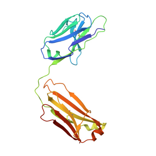
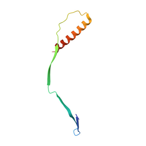
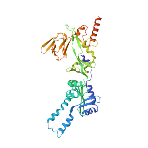
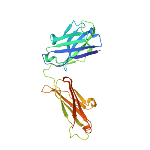
Improved epitope resolution of the prefusion trimer-specific antibody AM14 bound to the RSV F glycoprotein.

Harshbarger, W.Abeyrathne, P.D.Tian, S.Huang, Y.Chandramouli, S.Bottomley, M.J.Malito, E.

(2021) MAbs 13: 1955812-1955812

  • DOI: https://doi.org/10.1080/19420862.2021.1955812
  • Primary Citation of Related Structures:  
    7MMN, 7MPG

  • PubMed Abstract: 

    Respiratory syncytial virus (RSV) is the most common cause of acute lower respiratory tract infections resulting in medical intervention and hospitalizations during infancy and early childhood, and vaccination against RSV remains a public health priority. The RSV F glycoprotein is a major target of neutralizing antibodies, and the prefusion stabilized form of F (DS-Cav1) is under investigation as a vaccine antigen. AM14 is a human monoclonal antibody with the exclusive capacity of binding an epitope on prefusion F (PreF), which spans two F protomers. The quality of recognizing a trimer-specific epitope makes AM14 valuable for probing PreF-based immunogen conformation and functionality during vaccine production. Currently, only a low-resolution (5.5 Å) X-ray structure is available of the PreF-AM14 complex, revealing few reliable details of the interface. Here, we perform complementary structural studies using X-ray crystallography and cryo-electron microscopy (cryo-EM) to provide improved resolution structures at 3.6 Å and 3.4 Å resolutions, respectively. Both X-ray and cryo-EM structures provide clear side-chain densities, which allow for accurate mapping of the AM14 epitope on DS-Cav1. The structures help rationalize the molecular basis for AM14 loss of binding to RSV F monoclonal antibody-resistant mutants and reveal flexibility for the side chain of a key antigenic residue on PreF. This work provides the basis for a comprehensive understanding of RSV F trimer specificity with implications in vaccine design and quality assessment of PreF-based immunogens.


  • Organizational Affiliation

    GSK, Vaccine Design and Cellular Immunology, Rockville, MD, USA.


Macromolecules
Find similar proteins by:  (by identity cutoff)  |  3D Structure
Entity ID: 1
MoleculeChains Sequence LengthOrganismDetailsImage
AM14 Fab Light ChainA [auth L],
B [auth E],
C [auth G]
219Homo sapiensMutation(s): 0 
Entity Groups  
Sequence Clusters30% Identity50% Identity70% Identity90% Identity95% Identity100% Identity
Sequence Annotations
Expand
  • Reference Sequence
Find similar proteins by:  (by identity cutoff)  |  3D Structure
Entity ID: 2
MoleculeChains Sequence LengthOrganismDetailsImage
Fusion glycoprotein F2D [auth A],
F [auth C],
H [auth J]
72Human respiratory syncytial virus A2Mutation(s): 0 
UniProt
Find proteins for P03420 (Human respiratory syncytial virus A (strain A2))
Explore P03420 
Go to UniProtKB:  P03420
Entity Groups  
Sequence Clusters30% Identity50% Identity70% Identity90% Identity95% Identity100% Identity
UniProt GroupP03420
Sequence Annotations
Expand
  • Reference Sequence
Find similar proteins by:  (by identity cutoff)  |  3D Structure
Entity ID: 3
MoleculeChains Sequence LengthOrganismDetailsImage
Fusion glycoprotein F1,FibritinE [auth B],
G [auth I],
I [auth K]
414Human respiratory syncytial virus A2Tequatrovirus T4
This entity is chimeric
Mutation(s): 6 
UniProt
Find proteins for P03420 (Human respiratory syncytial virus A (strain A2))
Explore P03420 
Go to UniProtKB:  P03420
Entity Groups  
Sequence Clusters30% Identity50% Identity70% Identity90% Identity95% Identity100% Identity
UniProt GroupP03420
Sequence Annotations
Expand
  • Reference Sequence
Find similar proteins by:  (by identity cutoff)  |  3D Structure
Entity ID: 4
MoleculeChains Sequence LengthOrganismDetailsImage
AM14 Fab Heavy ChainJ [auth M],
K [auth H],
L [auth D]
244Homo sapiensMutation(s): 0 
Entity Groups  
Sequence Clusters30% Identity50% Identity70% Identity90% Identity95% Identity100% Identity
Sequence Annotations
Expand
  • Reference Sequence
Small Molecules
Ligands 2 Unique
IDChains Name / Formula / InChI Key2D Diagram3D Interactions
NAG
Query on NAG

Download Ideal Coordinates CCD File 
M [auth B]2-acetamido-2-deoxy-beta-D-glucopyranose
C8 H15 N O6
OVRNDRQMDRJTHS-FMDGEEDCSA-N
GOL
Query on GOL

Download Ideal Coordinates CCD File 
N [auth M]GLYCEROL
C3 H8 O3
PEDCQBHIVMGVHV-UHFFFAOYSA-N
Experimental Data & Validation

Experimental Data

  • Method: X-RAY DIFFRACTION
  • Resolution: 3.57 Å
  • R-Value Free: 0.245 
  • R-Value Work: 0.202 
  • R-Value Observed: 0.203 
  • Space Group: C 1 2 1
Unit Cell:
Length ( Å )Angle ( ˚ )
a = 223.423α = 90
b = 183.002β = 103.138
c = 135.785γ = 90
Software Package:
Software NamePurpose
HKL-3000data reduction
HKL-3000data scaling
PHENIXphasing
Cootmodel building
PHENIXrefinement
SERGUIdata collection

Structure Validation

View Full Validation Report


Ligand Structure Quality Assessment 


Entry History 

Deposition Data

Revision History  (Full details and data files)

  • Version 1.0: 2021-09-08
    Type: Initial release
  • Version 1.1: 2023-10-18
    Changes: Data collection, Refinement description