7LVZ

Crystal structure of ADO


Experimental Data Snapshot

  • Method: X-RAY DIFFRACTION
  • Resolution: 1.89 Å
  • R-Value Free: 0.228 
  • R-Value Work: 0.191 
  • R-Value Observed: 0.193 

wwPDB Validation   3D Report Full Report


This is version 1.2 of the entry. See complete history


Literature

The Crystal Structure of Cysteamine Dioxygenase Reveals the Origin of the Large Substrate Scope of This Vital Mammalian Enzyme.

Fernandez, R.L.Elmendorf, L.D.Smith, R.W.Bingman, C.A.Fox, B.G.Brunold, T.C.

(2021) Biochemistry 60: 3728-3737

  • DOI: https://doi.org/10.1021/acs.biochem.1c00463
  • Primary Citation of Related Structures:  
    7LVZ

  • PubMed Abstract: 

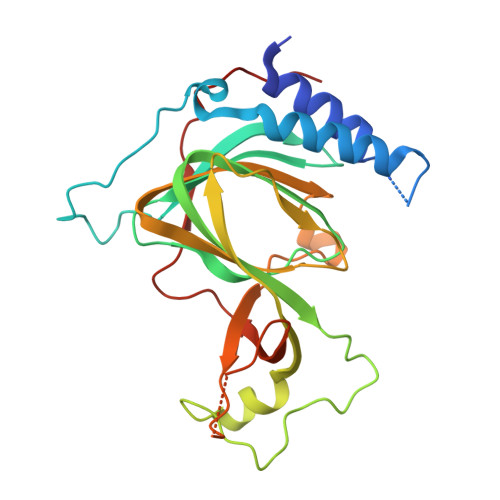
    We report the crystal structure of the mammalian non-heme iron enzyme cysteamine dioxygenase (ADO) at 1.9 Å resolution, which shows an Fe and three-histidine (3-His) active site situated at the end of a wide substrate access channel. The open approach to the active site is consistent with the recent discovery that ADO catalyzes not only the conversion of cysteamine to hypotaurine but also the oxidation of N-terminal cysteine (Nt-Cys) peptides to their corresponding sulfinic acids as part of the eukaryotic N-degron pathway. Whole-protein models of ADO in complex with either cysteamine or an Nt-Cys peptide, generated using molecular dynamics and quantum mechanics/molecular mechanics calculations, suggest occlusion of access to the active site by peptide substrate binding. This finding highlights the importance of a small tunnel that leads from the opposite face of the enzyme into the active site, providing a path through which co-substrate O 2 could access the Fe center. Intriguingly, the entrance to this tunnel is guarded by two Cys residues that may form a disulfide bond to regulate O 2 delivery in response to changes in the intracellular redox potential. Notably, the Cys and tyrosine residues shown to be capable of forming a cross-link in human ADO reside ∼7 Å from the iron center. As such, cross-link formation may not be structurally or functionally significant in ADO.


  • Organizational Affiliation

    Department of Chemistry, University of Wisconsin─Madison, Madison, Wisconsin 53706, United States.


Macromolecules
Find similar proteins by:  (by identity cutoff)  |  3D Structure
Entity ID: 1
MoleculeChains Sequence LengthOrganismDetailsImage
2-aminoethanethiol dioxygenase
A, B, C, D
256Mus musculusMutation(s): 0 
Gene Names: AdoGm237
EC: 1.13.11.19
UniProt
Find proteins for Q6PDY2 (Mus musculus)
Explore Q6PDY2 
Go to UniProtKB:  Q6PDY2
Entity Groups  
Sequence Clusters30% Identity50% Identity70% Identity90% Identity95% Identity100% Identity
UniProt GroupQ6PDY2
Sequence Annotations
Expand
  • Reference Sequence
Small Molecules
Ligands 3 Unique
IDChains Name / Formula / InChI Key2D Diagram3D Interactions
FE2 (Subject of Investigation/LOI)
Query on FE2

Download Ideal Coordinates CCD File 
E [auth A],
H [auth B],
K [auth C],
M [auth D]
FE (II) ION
Fe
CWYNVVGOOAEACU-UHFFFAOYSA-N
CL
Query on CL

Download Ideal Coordinates CCD File 
F [auth A],
I [auth B],
L [auth C],
N [auth D]
CHLORIDE ION
Cl
VEXZGXHMUGYJMC-UHFFFAOYSA-M
MG
Query on MG

Download Ideal Coordinates CCD File 
G [auth A],
J [auth B]
MAGNESIUM ION
Mg
JLVVSXFLKOJNIY-UHFFFAOYSA-N
Experimental Data & Validation

Experimental Data

  • Method: X-RAY DIFFRACTION
  • Resolution: 1.89 Å
  • R-Value Free: 0.228 
  • R-Value Work: 0.191 
  • R-Value Observed: 0.193 
  • Space Group: P 21 21 21
Unit Cell:
Length ( Å )Angle ( ˚ )
a = 54.296α = 90
b = 139.525β = 90
c = 142.007γ = 90
Software Package:
Software NamePurpose
PHENIXrefinement
PHASERphasing
autoPROCdata reduction
XDSdata scaling
Aimlessdata processing
STARANISOdata scaling

Structure Validation

View Full Validation Report


Entry History & Funding Information

Deposition Data


Funding OrganizationLocationGrant Number
National Institutes of Health/National Institute of General Medical Sciences (NIH/NIGMS)United States1R01GM117120-01A1
National Institutes of Health/National Institute of General Medical Sciences (NIH/NIGMS)United StatesT32GM008505

Revision History  (Full details and data files)

  • Version 1.0: 2022-01-05
    Type: Initial release
  • Version 1.1: 2024-02-28
    Changes: Data collection, Refinement description
  • Version 1.2: 2024-04-03
    Changes: Refinement description